Continue your surgical learning. One topic closer to your goals.
Textbook
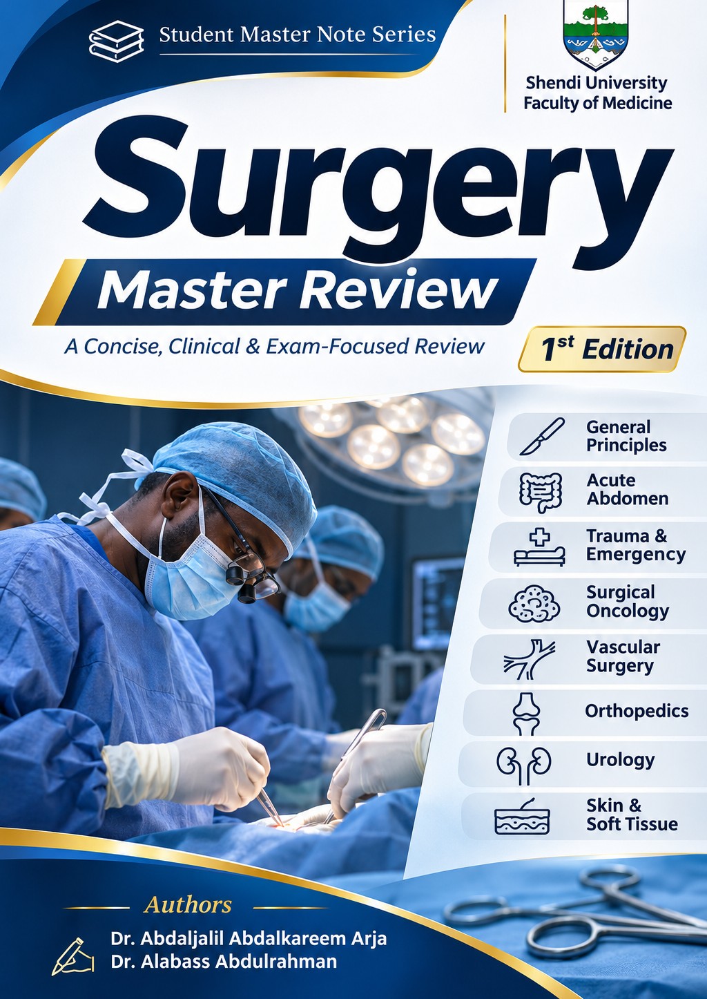
SurgeryMaster Review
Your surgical textbook
Clinical & exam focused
Start a study session
Study tools
My Notes & Highlights
Your progress
Recent Lectures
Explore the textbook
1 / 1
0:000:00
ابدأ رحلتك مع Surgery Master Review
مواضيع الفصل
تقدّمك
Contents
Student Master Note Series
عن المؤلفين
About the authors
Surgery Master Review
د. عبدالجليل ارجاد. العباس عبدالرحمن
خريجا كلية الطب — جامعة شندي مؤسِّسا سلسلة Student Master Note
من نحن
نحن خريجا كلية الطب بجامعة شندي، ومؤسِّسا سلسلة Student Master Note. عشنا سنوات الدراسة وسهر الليالي قبل الامتحانات، وأدركنا كيف يتشتّت وقت الطالب بين المراجع المطوّلة والملخّصات المتفرقة؛ فقرّرنا أن نُعِدّ المرجع الذي تمنّينا أن يكون بين أيدينا حين كنّا طلابًا. وكتاب Surgery Master Review هو كتاب الجراحة في السلسلة، بعد كتاب النساء والتوليد.
رحلة تجمعنا
جمعتنا الجامعة، وتوطّدت بيننا صداقة تمتدّ لأكثر من ثماني سنوات، أساسها الاحترام والتقدير المتبادل. نعتزّ بقدرات بعضنا، ونتكامل في أفكارنا وجهودنا، ونتشارك رؤية واحدة: أن نقدّم علمًا نافعًا يليق بطالب الطب ويعينه في رحلته.
رسالتنا
نؤمن بأن الفهم يسبق الحفظ، وبأن المعرفة تكتمل حين ترتبط بالممارسة. نسعى إلى تقريب العلم للطالب، وربط المراجع العالمية بواقع العمل الطبي في السودان، وتوجيه جهده إلى ما ينفعه في دراسته وامتحاناته.
منهجنا في العمل
نراجع المعلومة، وندقّق العبارة، ونعيد تنظيم المحتوى وتصميمه؛ إيمانًا بأن مشاركة العلم مسؤولية، وأن الطالب يستحق أدوات واضحة ومتقنة. ونرحّب بملاحظات زملائنا وطلابنا وأساتذتنا، ونتعلّم منها لنواصل التطوير.
نيّتنا
نسأل الله أن يكون هذا العمل خالصًا لوجهه الكريم، وعلمًا يُنتفع به، وصدقة جارية لنا ولوالدينا وأساتذتنا. فإن وجدت فيه نفعًا فلا تنسنا من دعوة صالحة، وإن وجدت خطأً فأعِنّا على تصحيحه.
Surgery Master Review · 1st edition · 2026Student Master Note Series
Student Master Note Series
عن هذه الطبعة
About this edition
Surgery Master Review
19فصلًا
76موضوعًا
2,793بطاقة مراجعة
1,002سؤالًا مع الشرح
تغطّي الطبعة الأولى مادة الجراحة لطلاب الطب في 19 فصلًا: المبادئ العامة، والبطن، والثدي والغدة الدرقية، والجلد والأنسجة الرخوة، والفحص السريري ومحطات OSCE، وأدوية الجراحة. ومع الكتاب بطاقات مراجعة لكل موضوع، وبنك أسئلة مع شرح الإجابات، ومراجعة سريعة تلخّص كل موضوع استعدادًا للامتحان.
المحتوى مبنيّ على الممارسة المعمول بها في المستشفيات السودانية، وتمّت مراجعته على المراجع الجراحية المعتمدة والإرشادات العلمية الحديثة.
في الطبعة الثانية بإذن الله: المسالك البولية، والأوعية الدموية، وجراحة الأطفال، وجراحة الصدر، وجراحة المخ والأعصاب.
إخلاء المسؤولية
هذا الكتاب وسيلة مراجعة لطلاب الطب، ولا يغني عن المراجع الأساسية ولا عن التدريب السريري ولا عن توجيه الأطباء المشرفين.
الطب علم متجدّد والخطأ وارد؛ لذا يجب التحقق من جرعات الأدوية وخطط العلاج من الإرشادات الحالية وبروتوكولات المستشفى قبل استخدامها مع المرضى. ولا يتحمّل المؤلفان أي مسؤولية عن ضرر أو خسارة تنتج عن استخدام هذا المحتوى.
جميع الحقوق محفوظة للمؤلفَين. الكتاب بكل محتواه — النصوص والجداول والرسومات، وبطاقات المراجعة، وبنك الأسئلة، والمراجعة السريعة، وتصميم التطبيق — ملكية فكرية خاصة بهما.
يُمنع دون إذن مكتوب من المؤلفَين
نسخ أي جزء من الكتاب أو طباعته أو تصويره أو نشر لقطات شاشة منه.
تحويله إلى ملف PDF أو أي صيغة أخرى.
مشاركته في مجموعات واتساب أو تيليجرام أو فيسبوك أو أي منصة أخرى.
بيعه أو توزيعه أو استخدامه في دورات أو ملخّصات تجارية.
الاشتراك شخصي: لطالب واحد على أجهزته فقط، ولا تجوز مشاركته مع شخص آخر؛ وأي مخالفة تؤدّي إلى إيقاف الاشتراك دون استرداد المبلغ. شراؤك للاشتراك الأصلي يدعم المؤلفَين ويضمن استمرار السلسلة وتحديثها.
شكر وتقدير
الحمد لله الذي بنعمته تتمّ الصالحات. نشكر والدينا على دعمهم الدائم، وأساتذتنا في كلية الطب بجامعة شندي الذين علّمونا الجراحة في العنابر وقاعات المحاضرات، والزملاء الذين أفادت ملخّصاتهم أجيالًا من الطلاب. وشكرًا لكل طالب يقرأ هذا الكتاب: ملاحظاتكم تجعل الطبعة القادمة أفضل — راسلونا على واتساب 0116984258.
Surgery Master Review · 1st edition · 2026Student Master Note Series
Student Master Note Series
فريق المراجعة
Reviewers
Surgery Master Review
بجهود مشتركة، أسهم فريق المراجعة في تدقيق المحتوى وتطويره، دعمًا لوضوح المادة العلمية وجودة تجربة التعلّم.
أريج يوسفنائبة الرئيسةجامعة أفريقيا العالميةInternational University of Africa
امنيه بكري عليرئيسة المراجعةجامعة الأحفاد للبناتAhfad University for Women
أعضاء فريق المراجعة
زياد نبيلعضو فريق المراجعةجامعة شنديShendi University
رحمه حسن حامدعضو فريق المراجعةجامعة شنديShendi University
سعاد عثمانعضو فريق المراجعةجامعة شنديShendi University
محمد عوض احمد عمرعضو فريق المراجعةجامعة الإمام المهديElimam Elmahdi University
احمد حمديعضو فريق المراجعةجامعة شنديShendi University
ملاك عبداللهعضو فريق المراجعةجامعة شنديShendi University
شكر وتقدير
نثمّن وقت المراجعين وجهودهم وملاحظاتهم البنّاءة، وإسهامهم في تحسين هذا العمل وتطويره. وتبقى مسؤولية الصياغة النهائية للمحتوى على عاتق المؤلفَين.
Surgery Master Review · 1st edition · 2026Student Master Note Series
Student Master Note Series
كيفية استخدام الكتاب والتطبيق
How to use the book
Surgery Master Review
دليل مبسّط للاستفادة من الكتاب وأدوات الدراسة.
1ابدأ بالفصل والموضوع
افتح «الفصول»، واختر الفصل ثم الموضوع، واقرأ صفحاته بالترتيب. يحفظ التطبيق آخر صفحة لتتابع منها.
2ثبّت فهمك بالأسئلة
من «التدريب» أنشئ اختبارًا: الوضع، ونوع الأسئلة، والفصول، والمستوى، والعدد؛ واقرأ الشرح بعد كل إجابة.
3راجع بالبطاقات
بطاقات اليوم من الرئيسية، أو بطاقات فصل أو موضوع؛ اقلب البطاقة ثم اختر: أعرفها أو أعدها لاحقًا.
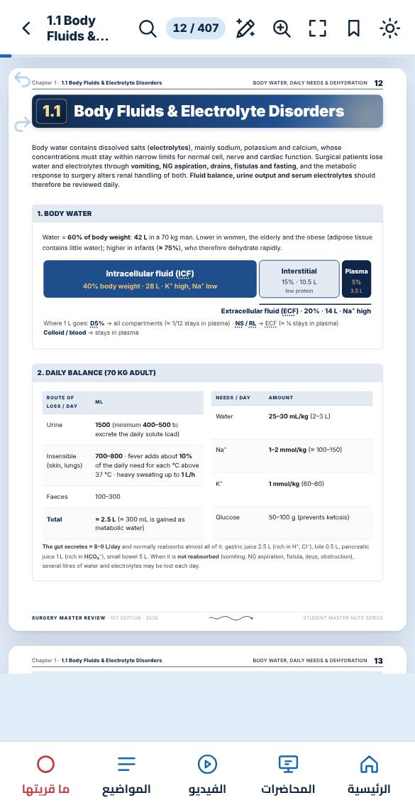
Body water contains dissolved salts (electrolytes), mainly sodium, potassium and calcium, whose concentrations must stay within narrow limits for normal cell, nerve and cardiac function. Surgical patients lose water and electrolytes through vomiting, NG aspiration, drains, fistulas and fasting, and the metabolic response to surgery alters renal handling of both. Fluid balance, urine output and serum electrolytes should therefore be reviewed daily.
1. Body water
Water = 60% of body weight: 42 L in a 70 kg man. Lower in women, the elderly and the obese (adipose tissue contains little water); higher in infants (≈ 75%), who therefore dehydrate rapidly.
2. Daily balance (70 kg adult)
Route of loss / day
mL
Urine
1500 (minimum 400–500 to excrete the daily solute load)
Insensible (skin, lungs)
700–800 · fever adds about 10% of the daily need for each °C above 37 °C · heavy sweating up to 1 L/h
Faeces
100–300
Total
≈ 2.5 L (≈ 300 mL is gained as metabolic water)
Needs / day
Amount
Water
25–30 mL/kg (2–3 L)
Na⁺
1–2 mmol/kg (≈ 100–150)
K⁺
1 mmol/kg (60–80)
Glucose
50–100 g (prevents ketosis)
The gut secretes ≈ 8–9 L/day and normally reabsorbs almost all of it: gastric juice 2.5 L (rich in H⁺, Cl⁻), bile 0.5 L, pancreatic juice 1 L (rich in HCO₃⁻), small bowel 5 L. When it is not reabsorbed (vomiting, NG aspiration, fistula, ileus, obstruction), several litres of water and electrolytes may be lost each day.
Surgery Master Review · 1st edition · 2026Student Master Note Series
↑ PCV (haemoconcentration) · urea rises more than creatinine · ↑ Na⁺ (if mostly water is lost) · concentrated urine (specific gravity > 1.020) with low urine Na⁺ (renal sodium conservation)
Best guide
Hourly urine output through a catheter: normal 0.5–1 mL/kg/h (≈ 30–50 mL/h in an adult). It reflects renal, and therefore organ, perfusion · central venous pressure in critically ill patients
Potassium (K⁺) determines the resting membrane potential of cardiac and skeletal muscle; both low and high levels cause arrhythmias. 98% of body K⁺ is intracellular, so any shift across cell membranes (insulin, alkalosis, acidosis, tissue injury) changes the serum level rapidly.
Loss: vomiting / NG suction, diarrhoea, fistula, villous adenoma (mucus rich in K⁺), diuretics · shift into cells: insulin, alkalosis · low Mg²⁺ · K⁺-free IV fluids for days
Not excreted: renal failure, ACE-I, spironolactone · released from cells: crush injury, burns, reperfusion of an ischaemic limb, acidosis · stored blood (> 20 mmol/L in an old unit) · false: haemolysed or delayed sample
Often none until arrhythmia or cardiac arrest; weakness
Surgery Master Review · 1st edition · 2026Student Master Note Series
Chapter 1 · 1.1 Body Fluids & Electrolyte DisordersPotassium14
6. ECG changes
ECG and potassium · low K⁺: flat T, ST ↓, U wave, long QT · high K⁺: tall tented T → flat P → wide QRS → sine wave → VF / asystole
7. Treatment
Treat low K⁺
Mild: oral KCl
IV: KCl 20 mmol (1 ampoule), always diluted, never a bolus
Rule of 4 (local): drip over ≥ 4 h · urine ≥ 40 mL/h (to ensure renal excretion) · ≤ 40 mmol per litre · on a ward no faster than 10 mmol/h
How much? Deficit (mmol) ≈ (4 − serum K⁺) × weight (kg) × 0.4, plus the daily need of 1 mmol/kg; this is only an estimate, because 98% of K⁺ is inside cells, so recheck K⁺ every 4–6 h and adjust
Not rising → give Mg²⁺; hypomagnesaemia causes renal K⁺ loss
Treat high K⁺: 3 steps
Protect the heart:Ca gluconate 10% 10 mL IV: stabilises the myocardium within minutes but does not lower K⁺
Shift K⁺ into cells:insulin 10 U + 50 mL 50% glucose (glucose prevents hypoglycaemia) · salbutamol neb · NaHCO₃
CCalcium gluconateBBicarbonate, β-agonist (salbutamol)IInsulinGGlucoseKK⁺-binding resin (calcium resonium)DROPDiuretics and dialysis
Exam trap
Never give KCl as an IV push: it causes cardiac arrest. In DKA and when starting insulin or feeding, K⁺ shifts into cells and falls rapidly: check it every 2–4 h. Blood from a long-stored unit, or releasing a tourniquet after long limb ischaemia, can raise K⁺ to a dangerous level.
Surgery Master Review · 1st edition · 2026Student Master Note Series
Na⁺ is the main extracellular cation and determines water distribution. An abnormal Na⁺ usually reflects a disorder of water balance: water excess → hyponatraemia; water deficit → hypernatraemia. The brain is mainly affected: cerebral oedema in hyponatraemia, cell shrinkage in hypernatraemia.
Hyponatraemia
Hypernatraemia
Level
Na⁺ < 135 mmol/L · fits usually < 120
Na⁺ > 145 mmol/L (normal 135–145)
Causes
Too much water: excess D5% / hypotonic fluid post-op (commonest in surgery, because post-operative ADH secretion causes water retention), SIADH (head injury, lung cancer, pain, drugs) · Na⁺ loss: vomiting, diarrhoea, fistula, diuretics, Addison's · pseudo: high glucose or lipids
Water loss more than Na⁺: inability to drink (elderly, unconscious or intubated patients), fever, sweating, diabetes insipidus (head injury), osmotic diuresis · excess saline
Features
Confusion, headache, nausea, fits, coma (brain swells)
Thirst, irritability, fits (brain shrinks)
Treatment
Water excess → restrict fluid · Na⁺ loss → normal saline · fits / coma → 3% saline in HDU. Rise per litre = (513 − serum Na⁺) ÷ (total body water + 1), where total body water = weight × 0.6 (men) or 0.5 (women, elderly)
Give water: oral, or D5% IV
Speed
Brain cells adapt over 24–48 h, so correct no faster than 8–10 mmol/L per 24 h: rapid correction of hyponatraemia → central pontine myelinolysis (brainstem damage) · rapid correction of hypernatraemia → cerebral oedema
Surgery Master Review · 1st edition · 2026Student Master Note Series
Ca²⁺ < 2.2 mmol/L (normal 2.2–2.6). Always correct for albumin, because half of serum Ca²⁺ is bound to it: corrected Ca (mg/dL) = measured Ca + 0.8 × (4 − albumin g/dL)
Ca²⁺ > 2.6 mmol/L
Causes
After thyroidectomy / parathyroidectomy (parathyroids injured or removed), acute pancreatitis (Ca²⁺ is used up in areas of fat necrosis), massive transfusion (citrate in stored blood binds Ca²⁺), low Mg²⁺, vitamin D deficiency
Symptomatic: Ca gluconate 10% 10 mL IV over 10 min, then an infusion (6–10 ampoules in 500 mL D5% over about 10 h) · oral Ca + vitamin D · correct Mg²⁺
IV normal saline 3–4 L/day first (corrects dehydration and promotes renal Ca²⁺ excretion) → then bisphosphonate (zoledronate 4 mg IV) · calcitonin · treat the cause
Mnemonic · CATs go numb (hypocalcaemia)
CConvulsionsAArrhythmias (long QT)TTetany: Chvostek and Trousseau signsnumbPerioral and finger numbness
Hypercalcaemia: "bones, stones, groans and psychic moans" (bone pain, renal stones, abdominal pain, confusion and depression).
10. Magnesium
Normal Mg²⁺ 0.7–1.0 mmol/L. Check it whenever K⁺ or Ca²⁺ will not correct.
Low: diarrhoea, fistula, NG losses, TPN, alcohol, diuretics, refeeding. Tremor, tetany, arrhythmia. It causes hypokalaemia and hypocalcaemia that do not respond to treatment (Mg²⁺ is required for renal K⁺ conservation and PTH secretion). Rx: MgSO₄ IV.
High: renal failure, excess Mg (antacids, MgSO₄ in eclampsia). Loss of reflexes first, then respiratory depression, heart block. Rx: stop Mg, Ca gluconate, dialysis.
Surgery Master Review · 1st edition · 2026Student Master Note Series
Chapter 1 · 1.1 Body Fluids & Electrolyte DisordersAcid–Base Balance17
Arterial pH is maintained between 7.35 and 7.45 by two systems: the lungs excrete CO₂ (acid) within minutes, and the kidneys retain or excrete bicarbonate over days. A primary disorder of ventilation is respiratory; a primary gain or loss of acid or bicarbonate is metabolic.
Step 1 pH · step 2 the value that explains the pH names the disorder (HCO₃⁻ moves with pH = metabolic; PCO₂ moves against pH = respiratory)
Step 3 · in metabolic acidosis, work out the anion gap = Na⁺ − (Cl⁻ + HCO₃⁻). It identifies the cause: high gap = an added acid has consumed HCO₃⁻ (lactate, ketones, uraemia) · normal gap = HCO₃⁻ has been lost (diarrhoea, fistula), with a compensatory rise in Cl⁻.
Normal anion gap = HCO₃⁻ lost from the gut or kidney: diarrhoea, fistula, ureterosigmoidostomy, renal tubular acidosis.
12. Acid–base disorders in surgical patients
Pyloric stenosis / GOO, repeated vomiting, NG suction
Loss of gastric acid (H⁺, Cl⁻) → hypochloraemic hypokalaemic metabolic alkalosis. Later paradoxical aciduria: in hypovolaemia the kidney reabsorbs Na⁺ in exchange for K⁺, and, once K⁺ is depleted, for H⁺, producing acid urine despite systemic alkalosis. Rx: NS + KCl (replaces Cl⁻ and K⁺); RL is inappropriate (lactate is metabolised to HCO₃⁻)
Diarrhoea, ileostomy, small bowel / pancreatic fistula
Normal anion-gap metabolic acidosis + hypokalaemia (loss of HCO₃⁻- and K⁺-rich fluid)
Shock, ischaemic bowel, sepsis
Lactic acidosis (high anion gap): anaerobic metabolism in hypoperfused tissue
Ureterosigmoidostomy
Hyperchloraemic acidosis, hypokalaemia
Pain, anxiety, early sepsis
Respiratory alkalosis (hyperventilation)
Massive transfusion
Later metabolic alkalosis (citrate → HCO₃⁻)
Surgery Master Review · 1st edition · 2026Student Master Note Series
Chapter 1 · 1.1 Body Fluids & Electrolyte DisordersAcid–Base Balance18
13. Compensation
The other system moves the pH back toward normal. The lungs act within minutes; the kidneys take 2–3 days. Compensation never overshoots the normal pH.
Primary disorder
Compensation
Metabolic acidosis (↓ HCO₃⁻)
Hyperventilation → ↓ PCO₂ (Kussmaul breathing in DKA)
Surgery Master Review · 1st edition · 2026Student Master Note Series
Chapter 1 · 1.2 IV Fluids & Fluid RegimensTypes of IV Fluid19
1.2IV Fluids & Fluid Regimens
Intravenous fluids should be prescribed like any drug: the correct fluid, volume and rate for a defined indication, reviewed daily. The indication is maintenance (the normal daily water and electrolytes of a patient who cannot eat or drink), replacement of ongoing losses, or resuscitation of a deficit or shock. Each fluid distributes to different body compartments (see 1.1), which determines the choice.
1. Crystalloids (mmol/L)
Crystalloids are aqueous solutions of small molecules (electrolytes, glucose). Saline solutions distribute through the extracellular fluid, so about a quarter remains intravascular; dextrose distributes through total body water once the glucose is metabolised.
Fluid
Na⁺
K⁺
Cl⁻
Other
Use
Normal saline 0.9%
154
–
154
9 g NaCl/L · osmolality 308 · pH ≈ 5.5 · 77 mmol Na⁺ per 500 mL
Resuscitation, vomiting / GOO, head injury
Ringer's lactate (Hartmann's)
131
5
111
Ca²⁺ 2 · lactate 29 (metabolised to HCO₃⁻ in the liver) · osmolality 278 · no glucose
Resuscitation, diarrhoea, fistula, burns
Dextrose 5% (D5%)
–
–
–
50 g glucose/L = 200 kcal/L; the glucose is metabolised, leaving free water
Maintenance water, hypernatraemia
DNS (5% dextrose in 0.9% saline)
154
–
154
Glucose 50 g/L + saline · hypertonic in the bottle
Maintenance when Na⁺ is also needed (local)
Surgery Master Review · 1st edition · 2026Student Master Note Series
Chapter 1 · 1.2 IV Fluids & Fluid RegimensTypes of IV Fluid20
Fluid
Na⁺
K⁺
Cl⁻
Other
Use
Dextrose-saline (4% / 0.18%)
30
–
30
"1/5 saline" + glucose
Maintenance
Hypertonic saline 3%
513
–
513
HDU only
Severe symptomatic ↓ Na⁺, raised ICP
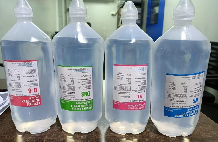
The 1-litre bottles on our wards · D-5 (5% dextrose) · DNS (0.9% saline + 5% dextrose) · RL (Ringer's lactate) · NS (0.9% saline). Label colours differ by maker: always read the label.
Colloids
Solutions of large molecules that do not cross the capillary wall and therefore remain intravascular, retaining water by oncotic pressure: albumin (4.5%, 20%), gelatins (Gelofusine, Haemaccel), dextran, starch (HES). They expand plasma volume faster but are more expensive and may cause anaphylaxis; dextran upsets cross-matching and clotting; starch harms the kidneys in sepsis. Guidelines recommend crystalloid first, as colloids show no survival benefit.
Blood
The fluid of choice for blood loss, as it restores oxygen-carrying capacity and clotting factors. ATLS: warmed crystalloid 1 L (children 20 mL/kg), then early blood if the response is transient. The former 3:1 rule (3 mL crystalloid per 1 mL blood lost) is abandoned: excess crystalloid causes haemodilution, hypothermia, coagulopathy and oedema.
2. Choosing the fluid
Gastric outlet obstruction, vomiting, NG losses
Normal saline + KCl: replaces lost Cl⁻ and K⁺. Not RL: its lactate adds HCO₃⁻ to an alkalosis
Diarrhoea, ileostomy, small-bowel fistula
Ringer's lactate: replaces lost HCO₃⁻ and K⁺
Haemorrhagic shock
RL / NS bolus while blood is prepared → blood
Head injury
Normal saline; avoid D5%: free water enters brain cells → cerebral oedema
Renal failure, hyperkalaemia
No K⁺-containing fluid (avoid RL, KCl)
Surgery Master Review · 1st edition · 2026Student Master Note Series
Chapter 1 · 1.2 IV Fluids & Fluid RegimensChoosing the Fluid & IV Access21
3. IV cannulae · a wider, shorter cannula gives faster flow
Flow is proportional to the fourth power of the radius (Poiseuille): doubling the radius increases flow 16-fold. A grey 16 G cannula infuses 1 L in about 6 min.
Colour · gauge · flow (mL/min) · red = trauma sizes: two orange / grey (14–16 G) · routine: green / pink · children: blue · neonates: yellow / violet
4. Venous access
Peripheral
First choice: forearm or back of hand, away from joints · re-site every 72–96 h or sooner if inflamed · two large-bore cannulae in shock · after 2–3 failed attempts, seek senior help
Central line
Internal jugular, subclavian or femoral · for CVP, TPN, vasopressors, irritant drugs, no peripheral veins · not the fastest route for volume · risks: pneumothorax, arterial puncture, air embolism, line sepsis, thrombosis
Intraosseous
Shocked child (or adult) when IV fails: needle into the marrow of the proximal tibia (flat anteromedial surface about 2 cm below the tibial tuberosity) or humeral head · the marrow drains into the venous circulation, so any fluid, blood or drug can be given
Cut-down
Long saphenous vein at the ankle, just in front of the medial malleolus
5. Complications of IV therapy
Thrombophlebitis (commonest: red, tender cord → remove cannula) · extravasation (KCl, calcium, 50% dextrose → skin necrosis) · infection, line sepsis · air embolism · fluid overload (pulmonary oedema, worst in the elderly and in heart or renal failure) · wrong fluid or electrolyte errors.
6. Prescribing IV fluids: three components
1 · MaintenanceNormal daily needs
2 · ReplacementMeasured ongoing losses
3 · ResuscitationExisting deficit / shock
Surgery Master Review · 1st edition · 2026Student Master Note Series
7. Maintenance: the 3-litre regimen (70 kg, local)
For an adult who cannot take oral fluids. One "drip" = 500 mL bottle; 6 bottles in 24 h, one pair every 8 h:
Hours
Bottles (2 × 500 mL)
KCl
0–8
D5% + D5%
1 ampoule (20 mmol)
8–16
NS + NS
1 ampoule (20 mmol)
16–24
D5% + D5%
1 ampoule (20 mmol)
Gives
3 L water · Na⁺ 154 mmol · K⁺ 60 mmol · glucose 100 g
Drip rate: an adult giving set gives 20 drops per mL (a paediatric burette 60). Drops/min = mL × 20 ÷ minutes: 500 mL over 4 h ≈ 42 drops/min.
NICE: 25–30 mL/kg/day water, 1 mmol/kg/day Na⁺, K⁺ and Cl⁻, 50–100 g glucose; less in the elderly and in heart or renal failure. Children (4-2-1 rule): 4 mL/kg/h for the first 10 kg + 2 mL/kg/h for the next 10 kg + 1 mL/kg/h for each kg above 20.
8. After surgery (local practice)
Day 1
3 L dextrose (D5% or dextrose-saline). No K⁺ (released from injured tissue) and little Na⁺ (retained under ADH and aldosterone)
Day 2
2 L D5% + 1 L NS
Day 3 on
Full regimen with KCl until eating and drinking; check U&E daily (too much D5% → hyponatraemia)
9. Replacement of ongoing losses
Measure every loss on the fluid balance chart and add it mL for mL, with a fluid of similar composition: NG aspirate / vomit → NS + KCl · diarrhoea, stoma, fistula → RL · fever: add about 10% of the daily need for each °C above 37 °C. In obstruction, ileus and peritonitis, litres of fluid are sequestered in the bowel and peritoneum (third-space loss): lost from the circulation but not recorded on the chart. Replace it with Ringer's lactate or normal saline (it has the composition of ECF), guided by pulse, BP and urine output. Daily weight helps: 1 kg ≈ 1 L of fluid.
10. Resuscitation
Worked example
70 kg, perforated DU, dry, pulse 110, postural drop → deficit ≈ 10% of body water (42 L) = ≈ 4 L of Ringer's lactate: half quickly, reassess, then the rest + maintenance; urine > 0.5 mL/kg/h.
Fluid challenge: 250–500 mL crystalloid over 15 min → reassess pulse, BP, urine, CVP. A small bolus tests fluid responsiveness without risking overload
Responds → repeat as needed · no response → think bleeding (blood, surgery) or another shock
Never give KCl as an IV push (cardiac arrest) or before the patient passes urine. A fit adult fasting only for a morning operation does not require IV fluids merely for being nil by mouth.
Surgery Master Review · 1st edition · 2026Student Master Note Series
Wound healing and immunity depend on adequate protein and energy. Many surgical patients are malnourished before admission (malignancy, dysphagia, vomiting, fistula), and surgery increases metabolic demand. Malnourished patients should be identified early and fed, by the enteral route whenever the gut is functional.
1. Importance of nutrition in surgery
Malnutrition → poor wound healing, anastomotic leak, wound dehiscence, infection and higher mortality. Surgery adds a catabolic state: stress hormones mobilise muscle protein for energy, reflected by ↑ urinary nitrogen (1 g nitrogen = 6.25 g protein).
2. Assessment
History
Weight loss > 10% in 3–6 months = severe · poor intake > 5–7 days · dysphagia, vomiting, GI cancer, fistula
Examination
BMI < 18.5 · muscle wasting · triceps skinfold (fat stores) · mid-arm muscle circumference (muscle protein) · hand-grip strength (muscle function: the first to fall and the first to recover)
Lab
Albumin < 30 g/L: a late and non-specific marker (half-life ≈ 20 days; also falls in sepsis and inflammation) · prealbumin (reflects recent change) · lymphocyte count
Needs
Energy 25–30 kcal/kg/day · protein 1–1.5 g/kg/day (more in sepsis and burns) · carbohydrate and protein give 4 kcal/g, fat 9 kcal/g
Best next step · "if the gut works, use it"
Severely malnourished before major elective surgery → feed for 7–14 days first, then operate: complications are reduced.
Surgery Master Review · 1st edition · 2026Student Master Note Series
Advantages: luminal nutrition preserves the mucosal barrier and reduces bacterial translocation; cheaper and safer than PN, with fewer infections
Practice: standard feed provides 1 kcal/mL · head elevated 30–45° during feeding to prevent aspiration
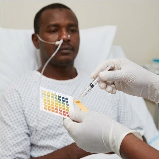
Complications:diarrhoea (commonest), aspiration, tube blockage or misplacement (confirm position before use: aspirate pH ≤ 5.5 or X-ray), metabolic upset
Contraindications:bowel obstruction, prolonged ileus, high-output proximal fistula, peritonitis, severe GI bleeding
4. Parenteral nutrition (PN / TPN)
Intravenous feeding, reserved for patients whose gut cannot be used; usually given as a single bag of about 2.5 L over 24 h.
Indications
Gut not usable: high-output enterocutaneous fistula, short bowel syndrome, prolonged ileus or obstruction, severe malabsorption
Route & content
Central vein (the hyperosmolar solution causes thrombophlebitis in peripheral veins): PICC or tunnelled Hickman line, with one lumen kept only for TPN (reduces infection) · glucose + lipid + amino acids + electrolytes, vitamins, trace elements
Complications
Line: sepsis (commonest serious), thrombosis, pneumothorax at insertion · Metabolic: hyperglycaemia, electrolyte upset, refeeding syndrome, fatty liver, cholestasis, gallstones (no enteral stimulus, so the gallbladder does not contract) · gut mucosal atrophy
Refeeding syndrome
Starvation depletes total-body phosphate, K⁺ and Mg²⁺. Feeding stimulates insulin release, which drives them into cells → severe hypophosphataemia (PO₄ < 0.5 mmol/L), hypokalaemia and hypomagnesaemia → arrhythmia, heart failure, death. Prevention: start low (≈ 10 kcal/kg/day) and increase gradually, give thiamine 200–300 mg daily before feeding (a cofactor for glucose metabolism), and check PO₄, K⁺, Mg²⁺ daily.
Surgery Master Review · 1st edition · 2026Student Master Note Series
Surgery Master Review · Perioperative Care2.1 Preoperative Assessment & Preparation27
2.1Preoperative Assessment & Preparation
Pre-operative assessment is the evaluation of a patient before an operation. Its purpose is to find and correct the conditions that make surgery and anaesthesia dangerous. Most deaths and complications around the time of surgery are caused by disease the patient already has, such as heart or lung disease, diabetes or kidney failure, rather than by the operation itself, and many of them can be prevented when they are recognised in advance.
Assessment before surgery
01Aims of pre-operative assessment
Confirm the indication for the operation.
Optimise co-morbid disease: heart, lungs, diabetes and kidneys.
Plan the anaesthetic, the blood products and, when needed, an ICU bed after surgery.
Obtain informed consent.
Prevent complications: surgical site infection, deep vein thrombosis (DVT) and pulmonary aspiration.
02History and examination
A careful history and examination is the best screening test before surgery. It finds more problems than any routine laboratory test, and it decides which investigations are needed.
Area
What to ask and look for
Heart and lungs
Angina, recent myocardial infarction (MI), heart failure, valve disease, hypertension, COPD, asthma and smoking. Elective surgery is delayed for 6 months after an MI, because the risk of a second infarction is highest in that period.
Exercise tolerance
A patient who can climb two flights of stairs without stopping has adequate heart and lung reserve for most operations.
Other diseases
Diabetes, kidney and liver disease, jaundice, epilepsy, sickle cell disease and pregnancy.
Medicines
Anticoagulants and antiplatelet drugs: warfarin, aspirin, clopidogrel.
Diabetic medicines (insulin, oral agents) and steroids.
Oral contraceptive pill (OCP) and hormone replacement therapy (HRT), which raise the risk of venous thromboembolism (VTE); anticonvulsants; diuretics, which lower K⁺.
Allergies
Penicillin and latex.
Previous anaesthesia
Difficult airway, malignant hyperthermia and suxamethonium apnoea. The family history is asked, because the last two are inherited.
Bleeding tendency
Easy bruising, prolonged bleeding after a dental extraction or an earlier operation, and a family history of bleeding. The history finds a patient who will bleed better than any laboratory test.
Surgery Master Review · 1st edition · 2026Student Master Note Series
Surgery Master Review · Perioperative Care2.1 Preoperative Assessment & Preparation28
03ASA physical status grade
The American Society of Anesthesiologists (ASA) grade describes how fit the patient is for anaesthesia. The letter E is added when the operation is an emergency (for example ASA III E).
Grade
Patient
Example
I
Normal healthy patient
Fit adult for a hernia repair
II
Mild systemic disease
Controlled hypertension or diabetes; a smoker
III
Severe systemic disease that limits activity
Stable angina; COPD with breathlessness
IV
Severe disease that is a constant threat to life
Recent MI, heart failure, sepsis
V
Moribund patient who will die without the operation
Ruptured abdominal aortic aneurysm in shock
VI
Brain-dead patient whose organs are being removed for donation
Organ donor
04Pre-operative investigations
Tests are chosen according to the patient's age, the diseases found in the history and the size of the operation.
Patient and operation
Investigations
Fit young patient, minor operation
No routine test is needed (NICE). In our hospitals a complete blood count (CBC) is the routine test, for example before an appendicectomy.
Major operation or older patient
CBC, urea and electrolytes (U&E) and random blood glucose. ECG over the age of 40–50 years or with heart disease. Chest X-ray with heart or lung disease and in the elderly.
Selected patients
Coagulation screen and liver function tests (LFT): anticoagulants, liver disease, jaundice.
Echocardiogram: a heart murmur or heart failure.
Pregnancy test (urine β-hCG): every woman of child-bearing age.
Sickling test (sodium metabisulphite slide, then haemoglobin electrophoresis if it is positive).
Viral screen: hepatitis B (HBV), hepatitis C (HCV) and HIV.
Group and save or cross-match when blood loss is expected.
Surgery Master Review · 1st edition · 2026Student Master Note Series
Surgery Master Review · Perioperative Care2.1 Preoperative Assessment & Preparation29
05Informed consent
Consent is valid only when the patient is competent, has been given enough information to decide, and decides freely.
Situation
Who gives consent
Competent adult (18 years or older)
The patient alone, even if the husband, wife or family disagree.
Child under 18 years
A parent or the legal guardian.
Emergency in an unconscious patient with no relative
The surgeon acts in the patient's best interest; the decision is documented by two doctors.
The surgeon explains the diagnosis, the operation, its benefits, its risks and complications, and the alternatives, including no treatment.
Preparing the patient
06Patients taking anticoagulant and antiplatelet drugs
Medicine
Management before and after surgery
Warfarin
The usual target INR is 2–3 (higher with a mitral or metallic heart valve).
Stop warfarin 5 days before surgery and operate when the INR is below 1.5.
Patients at high risk of clotting (metallic or mitral valve, recent VTE) need bridging: LMWH, with the last dose 24 hours before surgery, or a heparin infusion stopped 6 hours before. These short-acting drugs protect the patient until the operation.
Restart warfarin 12–24 hours after surgery.
Urgent reversal: vitamin K 10 mg IV with prothrombin complex concentrate (PCC) or fresh frozen plasma (FFP).
Aspirin and clopidogrel
Stop 7–10 days before surgery (local teaching: aspirin 2 weeks), because their effect lasts for the whole life of the platelet. Continue them after a recent coronary stent, because stopping risks stent thrombosis; the cardiologist is consulted.
Heparin
Unfractionated heparin is monitored by the APTT and reversed by protamine. LMWH causes less heparin-induced thrombocytopenia (HIT) and osteoporosis. LMWH is stopped 12 hours before spinal anaesthesia, because of the risk of a spinal haematoma.
Surgery Master Review · 1st edition · 2026Student Master Note Series
Surgery Master Review · Perioperative Care2.1 Preoperative Assessment & Preparation30
07Patients taking other regular medicines
Medicine
Management
Steroids (long-term)
Long-term steroids suppress the adrenal glands, which then cannot produce the cortisol needed for the stress of surgery. Steroid cover is given: hydrocortisone 100 mg IV at induction, then 50–100 mg every 6–8 hours, reduced gradually. Steroids also mask the signs of peritonitis, delay healing and increase infection.
OCP and HRT
Stop 4–6 weeks before major surgery, because they increase the risk of DVT.
Anticonvulsants
Continue; give them intravenously while the patient is nil by mouth.
Metformin
Omit on the day of surgery, because of the risk of lactic acidosis.
Smoking
Stop 6–8 weeks before surgery, so that the cilia of the airways and wound healing can recover.
08Patients with medical disease
Condition
Preparation
Diabetes
Place the patient first on the morning list, to shorten the fast and reduce the risk of hypoglycaemia.
Omit the oral agents and the morning insulin.
Give a sliding scale: IV insulin with dextrose and KCl, checking the blood glucose every 1–2 hours (target 6–10 mmol/L).
Obstructive jaundice
Bleeding: vitamin K 10 mg IV daily, with FFP if needed. Vitamin K is fat-soluble and is not absorbed when bile does not reach the gut.
Kidney failure (hepatorenal syndrome): IV fluids, with or without mannitol, and a urinary catheter to measure the urine output.
Make the patient euthyroid (carbimazole and propranolol, with or without Lugol's iodine) to prevent thyroid storm. Examine the vocal cords by indirect laryngoscopy, measure the baseline calcium, and take an X-ray of the neck and thoracic inlet to show tracheal deviation.
Gastric outlet obstruction
Correct the water, chloride and potassium loss with 0.9% saline and KCl. Pass a nasogastric (NG) tube and wash out the dilated stomach, and give nutritional support.
Colorectal surgery
Bowel preparation for operations on the left colon and rectum (oral laxative with or without an enema, and oral antibiotics). IV ceftriaxone and metronidazole at induction, repeated every 4 hours in a long operation. The stoma site is marked before surgery.
Sickle cell disease
Keep the patient hydrated, oxygenated and warm, because dehydration, hypoxia and cold cause sickling. Transfuse to a haemoglobin of about 10 g/dL, not higher, because thicker blood also causes sickling. Vaccinate at least 2 weeks before a splenectomy.
Anaemia and low platelets
The local minimum haemoglobin for elective surgery is 10 g/dL (guidelines accept 7–8 g/dL). The platelet count should be at least 50 000/mm³ for surgery.
Clinical caution
A patient on long-term steroids who becomes hypotensive soon after induction of anaesthesia may have an Addisonian (adrenal) crisis: give IV hydrocortisone at once.
Surgery Master Review · 1st edition · 2026Student Master Note Series
Surgery Master Review · Perioperative Care2.1 Preoperative Assessment & Preparation31
The day of surgery
09Prevention of venous thromboembolism
The risk of DVT and pulmonary embolism is estimated by adding the patient factors (age over 60, obesity, cancer, previous VTE, OCP or HRT, immobility, pregnancy) to the operation factors (major surgery, operations longer than 60 minutes, pelvic, hip or knee surgery). Scoring systems such as the Caprini score or the NICE assessment do this formally.
Risk
Prophylaxis
Low: young patient, minor operation
Early mobilisation
Moderate: major operation, age over 40, obesity
Add TED stockings, with or without LMWH
High: cancer, pelvic, hip or knee surgery, previous VTE
Add LMWH: enoxaparin 40 mg SC once daily, continued for 28 days after major cancer surgery
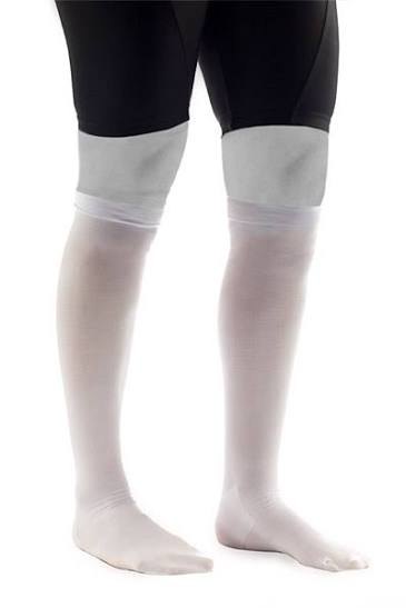
Figure 2.1.1 TED stockings give graduated compression from the toes to the thigh and speed up the venous return.
Enoxaparin doses. Prophylaxis: 40 mg SC once daily (20 mg when the creatinine clearance is below 30 mL/min). Treatment of DVT or pulmonary embolism: 1 mg/kg SC every 12 hours, or 1.5 mg/kg once daily.
10Preparation on the day of the operation
Fasting: 6 hours for food and 2 hours for clear fluids, to reduce the risk of pulmonary aspiration. In bowel obstruction an NG tube empties the stomach.
Mark the operation site, especially when the operation is on one side (left or right).
Remove hair with clippers just before the operation. A razor is never used the day before, because the small cuts it makes are colonised by bacteria and cause wound infection.
Give the prophylactic antibiotic within 60 minutes before the incision, so that the tissue level is high when bacteria enter the wound.
Order of the operating list: patients with latex allergy and diabetics first; dirty or infected cases last.
Complete the WHO surgical safety checklist.
Surgery Master Review · 1st edition · 2026Student Master Note Series
Surgery Master Review · Perioperative Care2.1 Preoperative Assessment & Preparation32
11WHO surgical safety checklist
Sign inbefore anaesthesia
Identity, site and procedure
Consent signed
Site marked
Allergies
Airway and aspiration risk
Expected blood loss; blood ready
Time outbefore the incision
Team members introduce themselves
Patient, site and operation confirmed
Antibiotic given within 60 minutes
Critical steps and blood loss
Imaging displayed
Equipment sterile
Sign outbefore leaving theatre
Operation recorded
Swab, needle and instrument counts correct
Specimen labelled
Equipment problems noted
Plan for recovery
The three pauses prevent operations on the wrong patient or the wrong side, swabs left in the patient and missed antibiotics.
12Sterilisation and disinfection
Sterilisation
Disinfection and antisepsis
Aim
Kills all organisms, including spores
Reduces the number of organisms; spores survive
Used on
Instruments, drapes and implants
Skin (antisepsis), surfaces and some endoscopes
Methods
Autoclave (steam under pressure, 121 °C for 15 minutes or 134 °C for 3 minutes): the best method for instruments.
Gamma radiation: disposable items.
Ethylene oxide or glutaraldehyde: heat-sensitive endoscopes.
Chlorhexidine, povidone-iodine and alcohol.
Boiling.
Exam point
Hand-washing is the single most important measure against the spread of infection in hospital. A competent adult alone gives consent: no relative can consent or refuse for them.
Surgery Master Review · 1st edition · 2026Student Master Note Series
Surgery Master Review · Perioperative Care2.2 Postoperative Complications33
2.2Postoperative Complications
Post-operative complications are problems that follow an operation. Each one tends to appear at a typical time after surgery, so the day since the operation is the most useful clue to the cause: a fever on day 1 has different causes from a fever on day 6. Early recognition and early treatment prevent most deaths.
General principles
01Classification of post-operative complications
By time after surgery
Immediate: within 24 hours.
Early: from the first days to 2 weeks.
Late: weeks to months.
By type
General: can follow any operation (chest, DVT, urinary, wound).
Specific: belong to the particular operation, for example hypocalcaemia after thyroidectomy.
Prevention
Optimise co-morbid disease before surgery.
Careful surgical technique.
Antibiotic and VTE prophylaxis.
Early mobilisation, chest physiotherapy, good analgesia and early feeding.
02Post-operative fever (the five Ws)
Fever is a temperature of 38 °C or more. A mild fever in the first 48 hours is often the normal inflammatory response to the operation; it needs no antibiotics if the patient is otherwise well.
Day
W
Usual cause
0–2
Wind
Atelectasis (the commonest cause) and pulmonary aspiration; also the normal inflammatory response, a transfusion or a drug reaction.
3–5
Water
Urinary tract infection from the catheter, chest infection, and phlebitis at the drip site.
5–7
Wound
Wound infection (rare before day 3) and anastomotic leak.
5–10
Walking and abscess
DVT and pulmonary embolism; an intra-abdominal abscess (pelvic or subphrenic), which gives a swinging fever.
Any day
Wonder drugs
Drug fever and blood transfusion. Malignant hyperthermia starts in theatre with muscle rigidity and a rising CO₂ and temperature; it is treated with dantrolene 2.5 mg/kg IV, and the mortality is about 10% even with treatment.
03Post-operative haemorrhage
Type
Time
Cause and management
Primary
During surgery
Controlled at the operation.
Reactionary
Within 24 hours
A ligature slips or a clot is dislodged as the blood pressure rises after anaesthesia and small vessels open (coughing, waking). Example: a neck haematoma after thyroidectomy, which can obstruct the airway; the wound is opened at the bedside, then the patient returns to theatre.
Secondary
7–14 days
Infection erodes the wall of a vessel, for example after haemorrhoidectomy or in an infected wound. Treatment: antibiotics, packing and re-exploration.
Surgery Master Review · 1st edition · 2026Student Master Note Series
Surgery Master Review · Perioperative Care2.2 Postoperative Complications34
04Respiratory complications
Complication
Features and management
Atelectasis
The commonest, on day 1–2 after upper abdominal or chest surgery. Pain stops deep breathing and coughing, so secretions block the small airways. The patient has fever, fast breathing and reduced breath sounds at the lung bases. An SpO₂ below 92% needs oxygen and arterial blood gases (ABG). Treatment: chest physiotherapy, deep breathing, analgesia and mobilisation.
Pneumonia
Day 3–5, with a productive cough and consolidation. Sputum culture and antibiotics.
Pulmonary aspiration
Vomiting under anaesthesia with a full stomach causes Mendelson's syndrome (acid pneumonitis). It usually affects the right lower lobe, because the right main bronchus is more vertical.
Pulmonary embolism
Day 5–10: sudden breathlessness, pleuritic chest pain, tachycardia and a falling SpO₂. Diagnosis by CT pulmonary angiogram; treatment with heparin.
The wound
05Wound complications
Complication
Features and management
Seroma and haematoma
Serum or blood collects under the wound, for example after mastectomy or hernia repair. It is aspirated or evacuated, because a haematoma is a good medium for bacteria.
Wound infection
Appears on day 3–7 with pain, redness and pus. The wound is opened (some sutures removed), drained, swabbed and dressed. Antibiotics are added only for surrounding cellulitis or systemic sepsis, because pus needs drainage, not antibiotics. It is the commonest complication after appendicectomy for a perforated appendix.
Incisional hernia
A late complication. Risk factors: wound infection, obesity and vertical midline incisions.
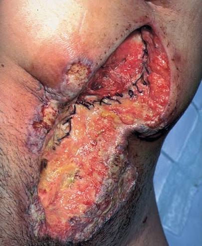
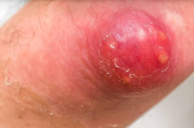
Figure 2.2.1 Wound infection: the sutures have been removed and the wound is left open to heal by granulation.Figure 2.2.2 Abscess: a red, hot, tense swelling with pus pointing to the surface; it needs drainage.
Surgery Master Review · 1st edition · 2026Student Master Note Series
Surgery Master Review · Perioperative Care2.2 Postoperative Complications35
06Burst abdomen (wound dehiscence)
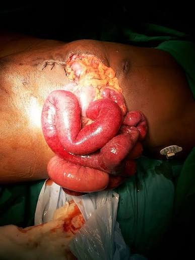
Figure 2.2.3 Burst abdomen: loops of bowel come out through a midline wound that has opened.
A burst abdomen usually occurs on day 7–10. The warning sign is a serosanguinous ("salmon-pink") discharge from the wound, followed by bowel appearing in the wound. The mortality is about 20%.
Causes
Technical failure of the closure: the most important cause.
Raised pressure in the abdomen: distension, coughing and vomiting.
Management
Reassure the patient and lay them flat.
Cover the bowel with sterile towels soaked in warm saline.
Give analgesia and IV fluids, pass an NG tube and start antibiotics.
Return to theatre for mass closure, with or without tension sutures.
Prevention
Mass closure with a non-absorbable or slowly absorbed suture.
Suture length at least 4 times the wound length.
Bites 1 cm from the edge and 1 cm apart.
Clinical caution
A salmon-pink discharge from an abdominal wound on day 7–10 means the deep layers have opened: inspect the wound and prepare for a burst abdomen.
Fistula and other complications
07Enterocutaneous fistula
A fistula is an abnormal communication between two epithelial surfaces; in an enterocutaneous fistula they are the bowel mucosa and the skin. The commonest cause is an anastomotic leak; others are Crohn's disease, cancer, radiation and a missed injury of the bowel.
A high-output fistula drains more than 500 mL a day, usually from the proximal small bowel. It causes dehydration, low K⁺, acidosis, malnutrition and excoriation of the skin. About two-thirds close without surgery, usually within about 6 weeks, once sepsis and nutrition are controlled.
Surgery Master Review · 1st edition · 2026Student Master Note Series
Surgery Master Review · Perioperative Care2.2 Postoperative Complications36
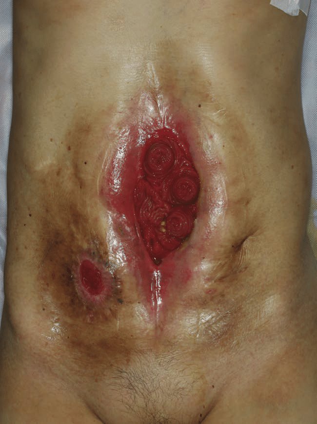
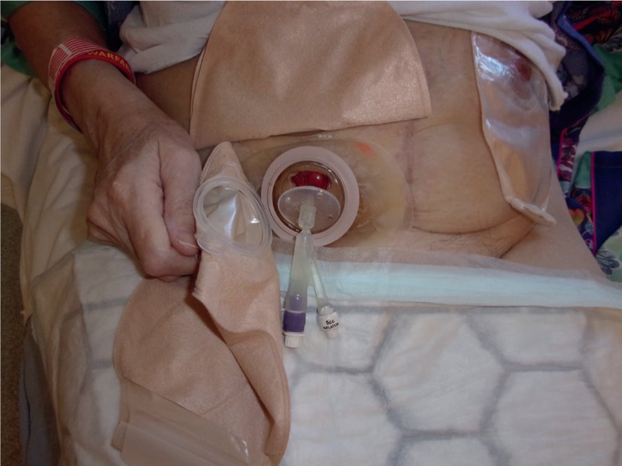
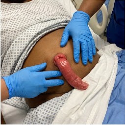
Figure 2.2.4 Intestinal fistulas opening into a broken-down laparotomy wound.Figure 2.2.5 The fistula output is collected in a stoma appliance, which measures the losses and protects the skin.
08Management of a fistula (SNAP)
S · SepsisDrain collections and give antibiotics
N · NutritionNil by mouth and TPN
A · AnatomyFistulogram and CT
P · PlanSurgery if it does not close
Step
What is done
Supportive care
Replace fluid and electrolytes, protect the skin with a stoma bag, and reduce the output with octreotide or a PPI.
Is it closing?
The daily output falls, the fever and tachycardia settle, and the weight and albumin rise. If the output stays high after about 6 weeks of good nutrition and sepsis control, look for a cause from the FRIENDS list below and plan surgery.
09Why a fistula does not close (FRIENDS)
Factor
Reason
F
Foreign body
A suture, mesh or drain keeps the track open.
R
Radiation
Irradiated tissue heals poorly.
I
Inflammatory bowel disease or infection
Crohn's disease or an abscess keeps the bowel diseased.
E
Epithelialised tract
A track lined by epithelium cannot close by itself.
N
Neoplasm
Cancer at the fistula.
D
Distal obstruction
Bowel content cannot pass onwards, so it leaves through the fistula.
S
Short track (under 2 cm) or steroids
A short track and steroids both prevent healing.
Surgery Master Review · 1st edition · 2026Student Master Note Series
Surgery Master Review · Perioperative Care2.2 Postoperative Complications37
10Deep vein thrombosis and pulmonary embolism
Deep vein thrombosis (DVT)
Pulmonary embolism (PE)
Time
Usually day 5–10 after surgery, often after a period of immobility.
Features
Calf pain, swelling, tenderness and warmth of one leg, sometimes with a low-grade fever. Many DVTs have no signs.
Sudden breathlessness, pleuritic chest pain, tachycardia, haemoptysis and a falling SpO₂; a large embolus causes collapse and shock.
Diagnosis
Clinical probability (Wells score), then compression duplex ultrasound. A normal D-dimer excludes DVT only when the probability is low; it is raised after any operation.
CT pulmonary angiogram. The ECG most often shows only sinus tachycardia; arterial blood gases show hypoxia.
Treatment
Therapeutic LMWH (enoxaparin 1 mg/kg every 12 hours), then warfarin or a direct oral anticoagulant for at least 3 months after a thrombosis provoked by surgery. A massive PE with shock is treated with thrombolysis.
11Post-operative oliguria
Oliguria is a urine output below 0.5 mL/kg/h (about 30 mL/h in an adult). After surgery the commonest cause is hypovolaemia (a pre-renal cause), which is corrected before the kidney is damaged.
Check the catheter first: a blocked or kinked catheter is flushed or changed.
Assess the fluid state: pulse, blood pressure, capillary refill, fluid balance chart, and losses in drains and the NG tube.
Give a fluid challenge of 250–500 mL of crystalloid over 15 minutes and watch the urine output.
If the urine does not increase, look for acute kidney injury: check urea, creatinine and K⁺, stop nephrotoxic drugs (NSAIDs, aminoglycosides), and ask for senior help.
12Other post-operative complications
Complication
Causes and management
Urinary retention
Pain, opioids, spinal anaesthesia, hernia or anal surgery, and benign prostatic hyperplasia (BPH). Treatment: a urinary catheter.
Paralytic ileus
Normal for 2–3 days after a laparotomy. When it lasts longer, look for a cause: low K⁺, uraemia, sepsis, anastomotic leak or opioids. Treatment: nil by mouth, NG tube and IV fluids.
Acute confusion
Hypoxia is excluded first, because it is common and quickly fatal. Other causes: sepsis, drugs (opioids), hypoglycaemia, low Na⁺, alcohol withdrawal and urinary retention.
Heart
A post-operative myocardial infarction is often silent (day 2–3), because analgesia masks the pain. In arrhythmias such as atrial fibrillation, check K⁺, Mg²⁺ and look for sepsis.
Others
Bleeding from a stress ulcer, pressure sores, acute kidney injury (AKI) from hypovolaemia, sepsis or drugs, and adhesions that cause obstruction later.
Surgery Master Review · 1st edition · 2026Student Master Note Series
Surgery Master Review · Perioperative Care2.2 Postoperative Complications38
13Anastomotic leak
An anastomotic leak is an escape of bowel contents through the join between two ends of bowel. It usually shows on day 5–7. The risk is highest after oesophageal and low rectal anastomoses, and is raised by malnutrition, a low albumin, steroids, shock, an emergency operation on unprepared bowel, and a join made under tension or with a poor blood supply.
Step
What is found or done
Features
Unexplained tachycardia (often the first sign), fever, new atrial fibrillation or confusion, pain more than expected, an ileus that does not settle, peritonitis, or bowel contents in a drain. The CRP keeps rising.
Investigation
CT of the abdomen with contrast (water-soluble contrast by mouth or rectum) shows free fluid, gas, a collection or contrast leaking out.
Management
Resuscitation, nil by mouth and broad-spectrum IV antibiotics. A small contained leak or abscess is drained percutaneously under CT or ultrasound guidance. Generalised peritonitis needs a relaparotomy: washout, and the ends are usually brought out as a stoma rather than joined again.
Clinical caution
A patient who develops tachycardia or atrial fibrillation on day 5–7 after a bowel anastomosis has a leak until it is proved otherwise.
14Post-operative pain and nausea
Good pain relief is part of treatment, not a comfort only: pain stops deep breathing, coughing and walking, and so leads to atelectasis, chest infection and DVT. Several drugs that act in different ways are combined (multimodal analgesia), so that smaller doses of each are needed.
Method
Use and cautions
Paracetamol
The base of every regimen: 1 g every 6 hours (maximum 4 g a day; less in liver disease and in patients under 50 kg).
NSAIDs
Diclofenac or ibuprofen add to paracetamol. Avoid them in kidney impairment, peptic ulcer, bleeding and asthma that worsens with aspirin.
Opioids
Morphine or pethidine for moderate to severe pain, given IV in small doses or by patient-controlled analgesia (PCA). Watch for respiratory depression, sedation, nausea, constipation and urinary retention; naloxone reverses them.
Local and regional
Wound infiltration with a local anaesthetic, nerve blocks and epidural analgesia, which gives the best relief after major abdominal surgery; hypotension is its main side effect.
Nausea and vomiting
Common after anaesthesia and opioids. Treat with ondansetron, metoclopramide or cyclizine; persistent vomiting needs a search for ileus or obstruction.
Exam point
The first thing to check in a confused patient after surgery is the oxygen saturation.
Surgery Master Review · 1st edition · 2026Student Master Note Series
Surgery Master Review · Perioperative Care2.3 Blood Transfusion39
2.3Blood Transfusion
Blood transfusion replaces what the patient has lost: red cells for oxygen delivery, and plasma or platelets for clotting. Every unit carries risks. The most dangerous is an ABO-incompatible transfusion, which is almost always caused by an error in identifying the patient or the unit. Blood is given only for a clear indication, and only the component that is missing is given.
Blood groups and compatibility
01Red cell compatibility
Recipient
O
A
B
AB
Can receive red cells of group
O
O, A
O, B
O, A, B, AB
Antibodies in the plasma
Anti-A, anti-B
Anti-B
Anti-A
None
O-negative red cells are the universal donor; AB-positive patients are the universal recipient.
Plasma works the other way: AB plasma is the universal plasma donor.
ABO: everyone has natural antibodies against the A or B antigens they lack, so red cells of the wrong ABO group are destroyed at once. This is the most dangerous mismatch.
Rh(D): an Rh-negative woman given Rh-positive blood forms anti-D antibodies, which can later destroy the red cells of an Rh-positive fetus (haemolytic disease of the newborn). Rh-negative girls and women of child-bearing age therefore receive Rh-negative blood.
02Preparing blood for the patient
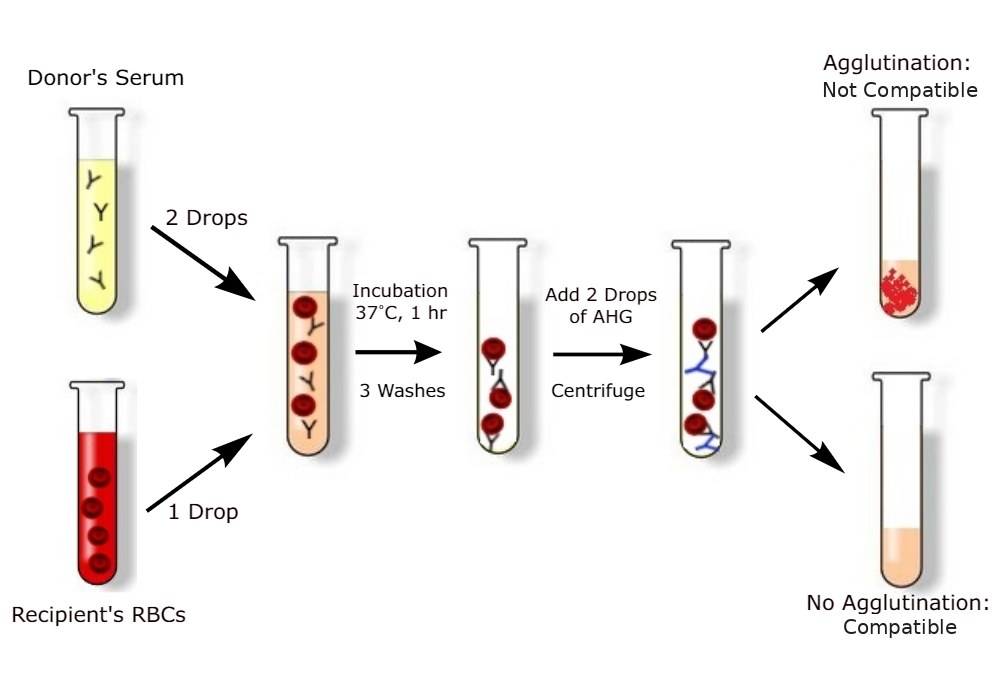
Figure 2.3.1 Cross-match: donor serum is mixed with the recipient's red cells; agglutination means the blood is not compatible.
Request
What the laboratory does
Group and save
Finds the blood group and screens the serum for antibodies; blood can then be issued quickly if it is needed.
Cross-match
Tests specific units against the patient's serum and reserves them for this patient.
Emergency with no time
O-negative red cells first, then group-specific blood.
Surgery Master Review · 1st edition · 2026Student Master Note Series
Surgery Master Review · Perioperative Care2.3 Blood Transfusion40
Blood products
03Blood products and their uses
Product
Storage
Given for
Fresh whole blood
2–6 °C
Acute massive bleeding when components are not available; common in our hospitals, where family members donate.
Packed red cells
2–6 °C, about 35 days
Anaemia and blood loss. They restore oxygen-carrying capacity, not clotting. One unit (from a donation of about 450 mL) raises the haemoglobin by about 1 g/dL.
Platelets
20–24 °C, shaken, 5 days
Bleeding with a low platelet count; before surgery when the count is below 50 000 (below 100 000 for brain or eye surgery); prophylaxis when it is below 10 000. One adult dose (4–6 pooled units or one apheresis unit) raises the count by about 20 000–40 000. Platelets are stored warm, because cold activates them.
Fresh frozen plasma (FFP)
−30 °C; thawed before use
Contains all the clotting factors (dose 15 mL/kg): coagulopathy, liver disease, DIC, massive transfusion, and reversal of warfarin together with vitamin K.
Cryoprecipitate
−30 °C
Rich in fibrinogen, factor VIII and von Willebrand factor (vWF): a fibrinogen below 1.5 g/L, and DIC.
Albumin and PCC
Room temperature
Albumin: hypoalbuminaemia and after paracentesis. Prothrombin complex concentrate (PCC): the fastest reversal of warfarin.
04When to transfuse red cells
Most patientsHb below 7 g/dL
Heart disease, the elderlyHb below 8 g/dL
Any HbActive bleeding or shock
Is it working? The Hb is checked again after the transfusion: each unit should raise it by about 1 g/dL, and the pulse should settle. If the Hb does not rise, suspect continuing bleeding or haemolysis and look for the source, rather than giving more blood blindly.
05Clotting tests
Test
What it measures and what prolongs it
PT and INR
The extrinsic pathway (factor VII). Prolonged by warfarin, vitamin K deficiency, obstructive jaundice and liver failure.
APTT
The intrinsic pathway. Prolonged by unfractionated heparin (UFH), haemophilia (factor VIII or IX deficiency) and DIC.
Platelet count and bleeding time
The platelet count shows thrombocytopenia. The bleeding time tests platelet function; it is prolonged by aspirin, uraemia and von Willebrand disease.
Safe for surgery
INR 1.5 or less, APTT ratio 1.5 or less, platelets at least 50 000 (at least 100 000 for brain or eye surgery) and fibrinogen at least 1.5 g/L.
Surgery Master Review · 1st edition · 2026Student Master Note Series
Surgery Master Review · Perioperative Care2.3 Blood Transfusion41
Giving blood and transfusion reactions
06Giving blood safely
Two people check the patient's identity, the blood group and the unit number at the bedside, because most deaths from transfusion are caused by clerical errors.
Record the baseline pulse, blood pressure and temperature.
Start slowly and watch closely for the first 15 minutes, when severe reactions appear.
A unit out of the fridge for more than 30 minutes is used or returned, and each unit is finished within 4 hours, because bacteria grow in warm blood.
Warm the blood when it is given rapidly.
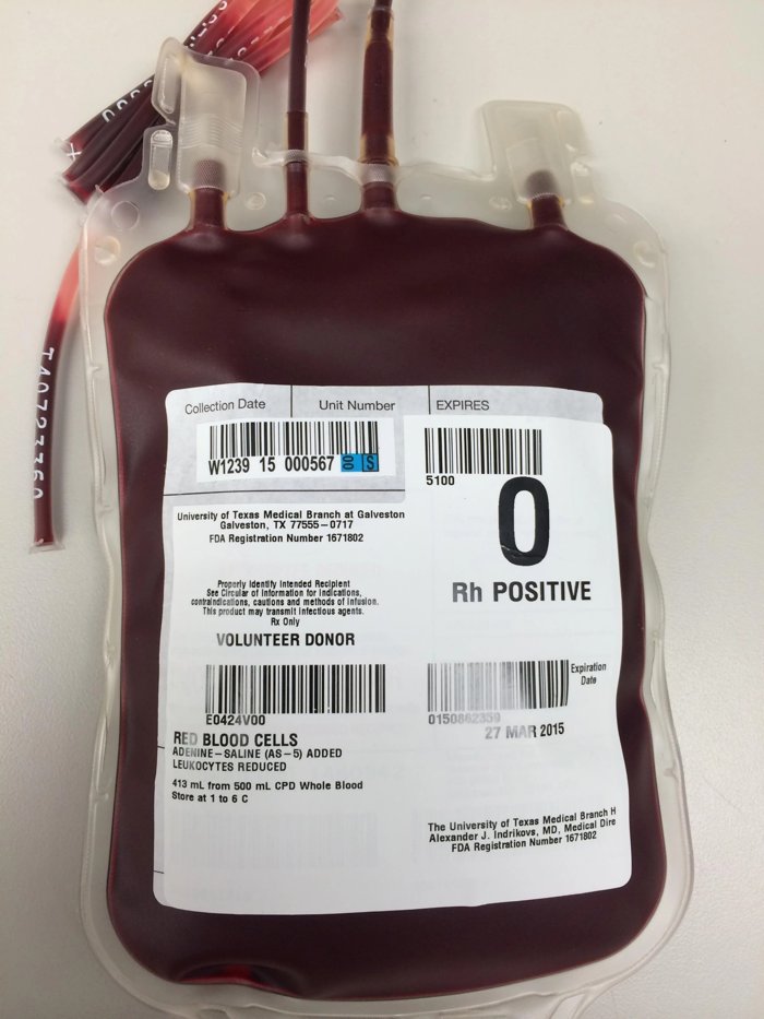
Figure 2.3.2 A unit of red cells: the blood group, unit number and expiry date are checked against the patient at the bedside.
07Transfusion reactions
Reaction
Picture
Action
Acute haemolytic
ABO incompatibility. Within minutes: fever, rigors, loin or chest pain, hypotension, haemoglobinuria, oliguria and DIC. Under general anaesthesia: unexplained hypotension and oozing from the wound.
Stop the transfusion, keep the vein open with saline, recheck the identity, send the bag and blood (direct antiglobulin test, Hb, bilirubin), give fluids to keep the urine flowing, and treat DIC.
Febrile non-haemolytic
The commonest reaction: fever and chills in a stable patient.
Slow or pause the transfusion; give paracetamol.
Allergic
Urticaria and itching. Anaphylaxis with bronchospasm and shock occurs in IgA deficiency.
Antihistamine. For anaphylaxis: stop, and give adrenaline IM.
Fluid overload (TACO)
Elderly patients and heart failure: breathlessness, crackles, a raised JVP and a raised blood pressure.
Stop, sit the patient up, give oxygen and furosemide.
Lung injury (TRALI)
Within 6 hours: breathlessness, hypoxia and bilateral infiltrates with a normal JVP (not overload).
Stop, give oxygen and ventilate if needed; no diuretic.
Delayed haemolytic
5–10 days later: jaundice and a falling Hb, from antibodies made after a previous transfusion or pregnancy.
Direct antiglobulin test and an antibody screen.
Infection
HBV, HCV, HIV, syphilis, malaria and CMV; bacterial contamination of platelets causes septic shock.
Screen the donors; culture the bag.
Surgery Master Review · 1st edition · 2026Student Master Note Series
Surgery Master Review · Perioperative Care2.3 Blood Transfusion42
Massive transfusion
08Definition and complications
Massive transfusion is the replacement of the patient's whole blood volume (in an adult about 70 mL/kg, about 5 L or 10 units) within 24 hours, of half of it within 3–4 hours, or of more than 4 units in 1 hour.
Complications
Hypothermia, the main cause of the coagulopathy: clotting enzymes fail below about 35 °C.
Hypocalcaemia: the citrate in stored blood binds calcium.
Hyperkalaemia: old red cells leak K⁺ (more than 20 mmol/L in an old unit).
Dilution of clotting factors and platelets.
Acidosis, later alkalosis, and ARDS.
Prevention
Warm all fluids and the patient.
Give red cells, FFP and platelets in a ratio of about 1 : 1 : 1.
Tranexamic acid 1 g IV in trauma, within 3 hours of the injury.
IV calcium, and stop the bleeding surgically.
09The lethal triad
HypothermiaCold fluids and an open abdomen slow the clotting enzymes
AcidosisShock causes lactic acidosis
CoagulopathySaline and red cells alone dilute the clotting factors
Each of the three worsens the other two, and together they cause more bleeding. The patient is kept warm, the bleeding is stopped early, and clotting products are given rather than saline alone.
Best next step
A trauma patient is still bleeding after 12 units: warm the patient, give calcium and clotting products, and pack the bleeding area (damage control surgery).
Exam traps
An acute haemolytic reaction causes fever, hypotension, haemoglobinuria and DIC, but not muscle rigidity.
Massive transfusion causes hypocalcaemia, never hypercalcaemia.
A patient on warfarin who is bleeding (for example with severe epistaxis) needs PCC or FFP with IV vitamin K; vitamin K alone is too slow.
A coagulopathy or a low platelet count contraindicates spinal anaesthesia.
Surgery Master Review · 1st edition · 2026Student Master Note Series
Student Master Note Series
SURGERYMaster Review
A concise revision guide for medical students · 1st edition · 2026
Tetanus & Gas GangreneClostridial infections · prophylaxis · treatment
55
Surgery Master Review1st edition · 2026
Surgery Master Review · Shock & Surgical Infection3.1 Shock44
3.1Shock
Shock is a state of inadequate tissue perfusion: the circulation fails to deliver enough oxygen to meet the needs of the cells. It is the commonest cause of death in surgical and trauma patients. The outcome depends on how early it is recognised, because the damage is reversible while the body is still compensating, but not once organs have begun to fail.
Recognising shock
01Definition and signs of hypoperfusion
Inadequate perfusion causes cellular hypoxia. The cells change to anaerobic metabolism and produce lactate; cell death and organ failure follow. The diagnosis rests on the signs of organ hypoperfusion, not on the blood pressure alone, because compensation keeps the blood pressure normal until late.
Organ
Sign of hypoperfusion
Skin
Cold, pale and clammy peripheries; capillary refill longer than 2 seconds.
Kidney
Oliguria: urine output below 0.5 mL/kg/h.
Brain
Anxiety, then confusion.
Blood
Lactate above 2 mmol/L. A shock index (heart rate divided by systolic blood pressure) above 1 also suggests shock while the blood pressure is still "normal".
02The body's response to shock
Compensation (still reversible)
Sympathetic drive: tachycardia.
Vasoconstriction: cold, pale skin.
Narrow pulse pressure, because the diastolic pressure rises.
Renin and antidiuretic hormone (ADH): oliguria and thirst.
Shock is a spiral, so it is treated in the compensated phase, before the blood pressure falls. The pulse pressure narrows early, because vasoconstriction raises the diastolic pressure while the stroke volume falls. Resuscitation aims for a mean arterial pressure (MAP) of at least 65 mmHg, the level needed to perfuse the kidneys and brain.
Surgery Master Review · 1st edition · 2026Student Master Note Series
Surgery Master Review · Shock & Surgical Infection3.1 Shock45
Spinal cord injury above T6, high spinal anaesthesia
↓
↓
↓
Warm, with bradycardia
Distributive shock is septic, anaphylactic and neurogenic shock: the vessels dilate and the systemic vascular resistance (SVR) falls, so the skin is warm. In the other types the sympathetic system constricts the vessels, so the skin is cold and the SVR high. Neurogenic shock causes bradycardia, because a cord injury above T6 cuts the sympathetic supply to the heart and leaves the vagus unopposed.
Distinguishing features
Distended neck veins after chest trauma: tension pneumothorax or cardiac tamponade. Distended veins with an MI on the ECG: cardiogenic shock.
Flat neck veins and a low cardiac output: hypovolaemic shock. The JVP is the best single sign that separates cardiogenic from hypovolaemic shock.
Warm hands with fever, for example in a diabetic with a gluteal abscess: septic shock.
Haemorrhagic shock
04Hidden sources of blood loss
Haemorrhage is the commonest cause of shock in surgical and trauma patients. A shocked trauma patient with no external bleeding is bleeding into one of five places: "on the floor and four more".
FloorExternal bleeding from wounds
ChestHaemothorax: chest X-ray
AbdomenLiver or spleen: FAST or CT
PelvisA pelvic fracture loses 3 L or more: pelvic X-ray and a pelvic binder
Long bonesA closed femoral fracture hides about 1.5 L in the thigh
Surgery Master Review · 1st edition · 2026Student Master Note Series
Surgery Master Review · Shock & Surgical Infection3.1 Shock46
05ATLS classes of haemorrhage
An adult's blood volume is about 70 mL/kg (about 5 L in a 70 kg man). The Advanced Trauma Life Support (ATLS) classes link the volume lost to the clinical signs, so that the loss can be estimated at the bedside before any investigation.
Class I
Class II
Class III
Class IV
Blood loss
Below 15% (below 750 mL)
15–30% (750–1500 mL)
30–40% (1500–2000 mL)
Above 40% (above 2000 mL)
Pulse
Normal
Above 100
Above 120
Above 140, thready
Systolic BP
Normal
Normal
Falls
Very low
Pulse pressure
Normal
Reduced
Reduced
Very reduced
Respiratory rate
14–20
20–30
30–40
Above 35
Urine (mL/h)
Above 30
20–30
5–15
Negligible
Mental state
Slightly anxious
Anxious
Confused
Lethargic
Treatment
Crystalloid
Crystalloid, with or without blood
Blood
Massive transfusion
Examples: after a road traffic accident, an anxious patient with a pulse of 110, BP 115/72 and a respiratory rate of 25 is in class II shock. A pale, restless patient with a pulse of 120 and BP 100/60 from a liver and spleen injury is in class III shock and needs blood.
Exam trap
Tachycardia is the first sign. The systolic blood pressure stays normal until about 30% of the blood volume is lost (class III), because vasoconstriction compensates. Young fit patients and pregnant women hide shock for longer, and elderly patients taking β-blockers may not become tachycardic.
06Management of haemorrhagic shock
Airway, breathing, circulation and high-flow oxygen. Stop the bleeding: direct pressure, splint fractures, a pelvic binder or a tourniquet.
Two large-bore cannulae. Send blood for group and cross-match, complete blood count (CBC), urea and electrolytes (U&E) and lactate.
A warmed crystalloid bolus, then blood early (red cells, FFP and platelets) and tranexamic acid. Large volumes of crystalloid dilute the clotting factors and worsen the bleeding.
Permissive hypotension until the bleeding is controlled (not in head injury), because a normal blood pressure can dislodge clots.
Find and control the source: chest X-ray, pelvic X-ray and FAST, then surgery or angio-embolisation.
Surgery Master Review · 1st edition · 2026Student Master Note Series
Surgery Master Review · Shock & Surgical Infection3.1 Shock47
07Response to the first fluid bolus
Rapid responderBecomes stable: has lost less than 20%. Observe, and image (CT) to find the injury.
Transient responderImproves, then the BP falls again within about 20 minutes: still bleeding. Give blood and operate.
Non-responderNo change: massive bleeding. Take the patient to theatre now.
08Monitoring resuscitation
Resuscitation is judged by the response of the patient, not by the volume of fluid given. The signs below show whether perfusion is being restored, when to stop giving fluid, and when the plan must change because the cause of shock has not been controlled.
Step
What is seen
First sign
The heart rate falls toward normal. It improves before the blood pressure, just as tachycardia was the first sign of shock.
Then
The blood pressure rises and the pulse pressure widens; capillary refill is under 2 seconds and the peripheries are warm; the mental state clears; the respiratory rate falls.
Best single guide
Urine output by catheter: at least 0.5 mL/kg/h in an adult (about 30–50 mL/h) and 1 mL/kg/h in a child, because it reflects the perfusion of the kidneys and so of the other organs.
Laboratory
The lactate falls (lactate clearance) and the base deficit improves. For example, urine 60 mL/h and lactate falling from 4 to 1.8 mmol/L mean that perfusion is restored.
Not improving
Tachycardia persists, the urine stays low and the lactate rises: ongoing bleeding or another cause of shock. Give blood and control the source, not more crystalloid. A normal blood pressure with a pulse of 125 and urine of 10 mL/h after 2 L of fluid still means shock.
When to stop fluid
The targets are reached and maintained: pulse, blood pressure, urine output and mental state normal, and the lactate falling.
There is no further response to a fluid challenge; more fluid only adds oedema.
Signs of fluid overload appear (basal crackles, a raised JVP, a falling SpO₂): stop and reassess.
Before the bleeding is controlled, aim only for permissive hypotension (systolic about 80–90 mmHg, radial pulse present). In head injury keep the systolic pressure at 100–110 mmHg or more to protect cerebral perfusion.
Surgery Master Review · 1st edition · 2026Student Master Note Series
Surgery Master Review · Shock & Surgical Infection3.1 Shock48
Septic shock
09Definitions
Sepsis is a life-threatening organ dysfunction caused by a dysregulated host response to infection. In surgical patients the source is usually in the abdomen (perforation, abscess, anastomotic leak, cholangitis), so antibiotics alone fail unless the source is drained or removed. Every hour of delay in giving antibiotics increases mortality.
Term
Definition
SIRS
Systemic inflammatory response syndrome: two or more of temperature above 38 °C or below 36 °C, heart rate above 90/min, respiratory rate above 20/min, and white cell count (WBC) above 12 000 or below 4000/mm³.
Sepsis
Infection with organ dysfunction. At the bedside, qSOFA of 2 or more: respiratory rate 22 or more, altered mental state, systolic BP 100 mmHg or less.
Septic shock
Sepsis that needs vasopressors to keep the MAP at 65 mmHg or more despite fluids, with a lactate above 2 mmol/L. A lactate above 4 mmol/L marks severe hypoperfusion and a high mortality.
Early (warm) phase
Warm, flushed skin and a bounding pulse; high cardiac output and low SVR; respiratory alkalosis, because the patient hyperventilates.
If the MAP stays below 65 mmHg after fluid, start noradrenaline, the vasopressor of first choice. The surgical key is source control within 6–12 hours: drain the abscess, remove dead tissue, and repair or exteriorise the perforation. A patient who is still hypotensive after fluids because of an abscess needs the abscess drained as well as noradrenaline.
11Monitoring the septic patient
Step
What is seen and done
First signs
The heart rate and respiratory rate fall, and the MAP holds at 65 mmHg or more.
Targets
MAP 65 mmHg or more, urine at least 0.5 mL/kg/h, lactate falling toward below 2 mmol/L (measured again within 2–4 hours), mental state clearing and capillary refill under 2 seconds.
Step down
Wean the noradrenaline once the MAP is stable. Narrow the antibiotic to the culture result at 48–72 hours, and stop it after about 5–7 days once the source is controlled and the fever and WBC have settled.
Failure: change the plan
A rising lactate or vasopressor dose, or fever and a high WBC that persist after 48–72 hours, mean an uncontrolled source (a residual abscess or a leak). Repeat the imaging (ultrasound or CT) and drain or re-operate; review the antibiotic and consider resistant organisms or fungi. Example: fever and a rising lactate on day 5 after an anastomosis despite antibiotics suggest a leak: CT, then drainage or re-operation.
Surgery Master Review · 1st edition · 2026Student Master Note Series
Surgery Master Review · Shock & Surgical Infection3.1 Shock49
The other types of shock
12Treatment of the other types
These types of shock are less common on surgical wards, but each has a specific treatment that reverses it quickly, so the cause must be recognised rather than treated with fluid alone.
Type
Typical picture
Treatment
Anaphylactic
Wheeze, cyanosis and a BP of 70/30 minutes after an IV antibiotic.
Stop the trigger; airway and high-flow oxygen; lay the patient flat with the legs raised.
Adrenaline 0.5 mg IM (0.5 mL of 1:1000) into the anterolateral thigh; repeat after 5 minutes if there is no improvement.
IV crystalloid bolus; then chlorphenamine and hydrocortisone, which act slowly and prevent recurrence.
Neurogenic
After a road traffic accident: BP 80/30, pulse 50, warm skin and paralysed legs.
IV fluids with care, a vasopressor, and atropine for the bradycardia; immobilise the spine.
Cardiogenic
Shock with distended neck veins and an MI on the ECG.
Treat the cause (percutaneous coronary intervention (PCI) for an MI, correct the arrhythmia) and give inotropes (dobutamine). Avoid fluid overload, because the failing heart cannot pump the extra volume and pulmonary oedema follows.
Obstructive
A stab wound of the chest with a low BP, raised JVP and muffled heart sounds (tamponade).
Relieve the obstruction: needle decompression then a chest drain for a tension pneumothorax; pericardiocentesis for tamponade; thrombolysis for a massive PE.
Anaphylaxis: is the treatment working?
First signs: the wheeze and stridor settle, the blood pressure rises and the pulse slows.
No improvement 5 minutes after adrenaline: repeat the IM dose. After two doses with no response, call the ICU for an adrenaline infusion.
Observe for 6–12 hours even after full recovery, because a biphasic reaction can recur.
Exam traps
Bleeding of more than 1 L causes reduced renal blood flow, a rise in the platelet count, metabolic acidosis and a low CVP; it does not cause alkalosis or a raised CVP.
The first response in septic shock is tachycardia, and septic shock is a state of low peripheral resistance.
High-dose steroids and anti-endotoxin antibodies do not reduce deaths from sepsis; early antibiotics and source control do. The commonest cause of death in severe acute pancreatitis is sepsis with multi-organ failure.
Sudden septic shock years after a splenectomy is overwhelming post-splenectomy infection (OPSI), usually by pneumococcus, H. influenzae or meningococcus. Vaccinate at least 2 weeks before an elective splenectomy.
In anaphylaxis outside the ICU, adrenaline is given IM, not as an IV bolus.
Surgery Master Review · 1st edition · 2026Student Master Note Series
Wound healing is the restoration of tissue continuity after an injury or an incision. Every wound heals in the same sequence of inflammation, proliferation and remodelling, but how fast and how well it heals depends on the blood supply, on infection and on the general condition of the patient. A wound that fails to heal leads to infection, dehiscence, an incisional hernia or an ugly scar.
Wound healing
01Types of wound healing
Type
How the wound heals
Example
Primary intention
Clean edges are brought together and unite without breakdown, leaving a minimal scar. The epithelium covers a sutured wound in 24–48 hours.
Surgical incision; a wound closed with a subcuticular suture
Secondary intention
The wound is left open. The cavity fills with granulation tissue, then contracts (myofibroblasts) and is covered by epithelium. Healing is slower and the scar bigger.
Drained abscess cavity packed and left open, infected wound, ulcer
Tertiary (delayed primary closure)
A contaminated wound is left open for 3–5 days, then closed once it is clean, because closing a dirty wound traps bacteria and causes an abscess.
Dirty traumatic wound; skin wound of a perforated appendix
02Phases of healing
InflammatoryDay 0–4 Clot, then neutrophils and macrophages clear bacteria and dead tissue
ProliferativeDay 4 to week 3 Fibroblasts lay down type III collagen; granulation tissue, new vessels and new epithelium
RemodellingWeek 3 to 1 year Type III collagen is replaced by type I collagen and the scar matures
On day 1 the strength of a wound comes only from the sutures. The scar regains about 70% of the strength of normal skin by 6 weeks, and never more than about 80%, because remodelled collagen is never as well organised as normal skin.
03Granulation tissue
Healthy
Unhealthy
Appearance
Red, granular, and bleeds on touch
Pale, flat and sloughy
Meaning
Good blood supply: the wound is ready for a skin graft
Ischaemia or infection: the cause is treated first
Surgery Master Review · 1st edition · 2026Student Master Note Series
Sutures on the face are removed early because its rich blood supply heals the wound fast, and early removal avoids track marks.
Suture
Examples
Used for
Absorbable
Vicryl, PDS, catgut
Deep layers, bowel and subcuticular skin closure; they need no removal.
Non-absorbable
Nylon, Prolene, silk
Skin, hernia repair and vessels, where the strength must last.
05Factors that delay healing
Local
Infection: the commonest cause.
Poor blood supply and ischaemia.
Haematoma and dead space.
Foreign body and necrotic tissue.
Tension and movement.
Radiation.
General
Old age.
Malnutrition: low albumin, vitamin C and zinc.
Diabetes, anaemia, jaundice and uraemia.
Steroids and chemotherapy.
Cancer, smoking and obesity.
Memory aid
DIDN'T HEAL: Diabetes, Infection, Drugs (steroids, chemotherapy), Nutritional deficiency (protein, vitamin C, zinc), Tissue necrosis and foreign body, Hypoxia and ischaemia, Excessive tension, Another wound or repeated trauma, Low temperature. Antibiotics do not delay healing.
Surgical site infection
06Surgical site infection (SSI)
A surgical site infection is an infection of the wound or of the operated organ within 30 days of surgery, or within 1 year when an implant (mesh, prosthesis) is left in place. It is the commonest hospital-acquired infection in surgical patients, and its risk depends mainly on how contaminated the wound is. It presents with pain, redness, swelling, pus and fever, usually on days 5–7; its treatment is described in 2.2.
Depth
Superficial: skin and subcutaneous fat.
Deep: fascia and muscle.
Organ or space: for example an intra-abdominal abscess.
Organisms
Staphylococcus aureus: the commonest, in clean wounds.
E. coli and Bacteroides: bowel surgery.
Streptococcus pyogenes: spreading cellulitis.
Pseudomonas: blue-green pus.
Distinguishing features
A wound infection on day 1–2 with severe pain or bronze skin is not a routine SSI: think of clostridia (gas gangrene, 3.3) or Streptococcus, which spread fast and need urgent surgery.
Surgery Master Review · 1st edition · 2026Student Master Note Series
Diabetes, obesity, malnutrition, smoking, steroids, old age, and a long pre-operative stay, in which the skin is colonised by hospital organisms.
Operation
Contamination (the wound class) and a long operation.
Shaving the night before: the small skin cuts are colonised overnight, so hair is clipped just before surgery instead.
Haematoma, dead tissue and drains; hypothermia and poor technique.
An upper respiratory tract infection is not a risk factor for SSI.
08Wound classification (CDC)
Class
Definition
Examples
SSI rate
Clean
No hollow viscus opened and no inflammation
Hernia repair (with or without mesh), thyroidectomy, varicose veins, excision of a fibroadenoma
Below 2%
Clean-contaminated
Gastrointestinal, biliary, urinary or respiratory tract opened under control
Cholecystectomy (open or laparoscopic), elective colectomy, appendicectomy for simple appendicitis
Below 10%
Contaminated
Spillage from the gut, acute inflammation without pus, a fresh trauma wound
Gross spillage at colectomy, an open fracture less than 4 hours old
15–20%
Dirty
Pus or a perforation already present, an old trauma wound
Perforated appendix with an abscess, faecal peritonitis
Above 30%
Exam trap
Clean-contaminated comes before contaminated: the bowel is opened, but under control. Some local notes swap them; learn the order clean, clean-contaminated, contaminated, dirty.
09Antibiotic prophylaxis
Prophylactic antibiotics aim for a high concentration of the drug in the tissues at the moment the wound is contaminated, so that bacteria are killed before they can multiply. They reduce SSI only when they are given at the right time; long courses add resistance and C. difficile colitis without any benefit.
Rule
Who needs it
Clean-contaminated and contaminated operations.
Clean operations with a prosthesis (mesh, joint, graft), such as an inguinal hernia repair with mesh, because infection of an implant usually means removing it.
Dirty wounds, such as a perforated appendix with pus, need a treatment course, not one prophylactic dose.
When
IV at induction (within 60 minutes before the incision) as a single dose. It is repeated in operations longer than about 4 hours or with blood loss above 1.5 L, because the tissue level falls.
Which drug
Clean operations: cefazolin. Bowel and appendix (our practice): ceftriaxone 1 g with metronidazole 500 mg IV.
Surgery Master Review · 1st edition · 2026Student Master Note Series
Ceftazidime, piperacillin-tazobactam or ciprofloxacin
Clostridium difficile
Oral vancomycin or metronidazole
Mixed abdominal sepsis (our practice)
Ceftriaxone with metronidazole; meropenem when severe
11Is the antibiotic treatment working?
Step
What is seen and done
First signs
The fever and pulse settle, and the pain and redness recede within 48–72 hours. The edge of a cellulitis is marked with a pen to see whether it spreads.
Then
The WBC and C-reactive protein (CRP) fall; the patient eats and walks.
Step down and stop
Change from IV to oral once the patient has been afebrile for 24–48 hours and is eating. Narrow the drug to the culture result, and stop after 5–7 days for most surgical infections once the source is controlled.
Failure: change the plan
Fever after 72 hours means undrained pus (an abscess, a collection or a leak) until proved otherwise. Examine the wound, arrange ultrasound or CT and drain; then review the culture and the drug.
12Infection control
Hand-washing before and after every patient: the single most important measure against cross-infection in hospital.
Sterile technique and safe disposal of sharps.
Universal precautions for every patient, whether HIV-positive or not; an HIV-positive patient with a perianal abscess has the usual incision and drainage.
Every surgeon needs hepatitis B (HBV) vaccination.
Best next step
Needle-stick injury: wash under running water and let the wound bleed, and report the injury. Test the source patient and the staff member. Give HIV post-exposure prophylaxis (PEP) when the risk is high: best within 2 hours, and no later than 72 hours. Give an HBV booster or immunoglobulin to a worker who is not immune. The risk of transmission from an infected source is about 30% for HBV (non-immune), 3% for hepatitis C (HCV) and 0.3% for HIV, so a vaccinated surgeon's main risk is hepatitis C.
Surgery Master Review · 1st edition · 2026Student Master Note Series
An abscess is a collection of pus walled off by granulation tissue. The wall protects the body but also keeps antibiotics out, which is why pus must be drained. Most superficial abscesses are caused by Staphylococcus aureus.
It is a hot, red, tender, fluctuant swelling with a swinging fever. Fluctuation may be absent in a deep abscess (breast, buttock), where ultrasound or needle aspiration confirms the pus.
A cold abscess has no heat or redness. It is caused by tuberculosis, for example in the neck or the psoas: a painless, cold, fluctuant neck swelling with night sweats.
Treatment: "where there is pus, let it out"
Incision and drainage under anaesthesia: break down the loculi, wash out, and leave the cavity open or packed.
Antibiotics alone do not cure an abscess, because they cannot pass through the wall. Add them for cellulitis, diabetes or sepsis.
Send the pus for culture and check the blood glucose.
Working: the pain eases within 24 hours and the fever settles. A persistent fever means a loculus has been left undrained.
14Common skin and hospital-acquired infections
Infection
Features and treatment
Boil (furuncle)
Staphylococcal infection of one hair follicle: warm compresses, and drainage if it points. Boils that recur every 2–3 weeks suggest diabetes, so the glucose is checked.
Carbuncle
Infection of many adjacent hair follicles with sloughing and multiple openings (cribriform), usually on the back of the neck in a diabetic. Treatment: drainage, antibiotics and control of the blood glucose.
Felon (pulp space infection)
An abscess of the fingertip pulp with throbbing pain. It is drained early, because the pressure in the closed pulp space can cause osteomyelitis of the terminal phalanx.
Lymphangitis
Red streaks up the limb from a wound (for example a thorn in the foot), with tender lymph nodes and fever. It is caused by Streptococcus pyogenes: admit for IV penicillin and elevate the limb.
C. difficile colitis
Diarrhoea with pseudomembranes after antibiotics, classically clindamycin and cephalosporins. Diagnosis: stool toxin assay. Treatment: stop the antibiotic, isolate the patient, and give oral vancomycin 125 mg every 6 hours for 10 days.
MRSA
Isolate the patient, hand hygiene, screening and decolonisation; treatment with vancomycin.
Covered elsewhere: wound infection, burst abdomen and fistula after surgery in 2.2; cellulitis, necrotising fasciitis and Fournier's gangrene in Skin & Soft Tissue; gas gangrene and tetanus in 3.3.
Surgery Master Review · 1st edition · 2026Student Master Note Series
Surgery Master Review · Shock & Surgical Infection3.3 Tetanus & Gas Gangrene55
3.3Tetanus & Gas Gangrene
Tetanus is a disease of the nervous system caused by the toxin of Clostridium tetani, which grows in a contaminated wound. It is still common in Sudan, especially after farm injuries and in newborns delivered at home, and its mortality reaches 60% where intensive care is limited. It is entirely preventable by vaccination and proper wound care.
Tetanus
01Cause and mechanism
SporesAnaerobic, Gram-positive, spore-forming "drumstick" bacillus from soil and faeces
GerminationThe spores grow in dead, low-oxygen tissue and release tetanospasmin
Toxin actionIt travels up the nerves and blocks the inhibitory neurones (GABA, glycine)
With the inhibitory neurones blocked, muscles contract without opposition (spasms) and the sympathetic system overacts. The incubation period, from the wound to the first symptom, is 3–21 days, usually 7–10 days; a shorter incubation means more severe disease.
02Clinical features
Trismus (lockjaw): usually the first sign.
Risus sardonicus: a fixed grin.
Stiffness of the neck and back, leading to opisthotonus (an arched back).
Reflex spasms triggered by noise, light or touch; spasm of the larynx causes asphyxia.
The patient stays fully conscious.
Autonomic storm: sweating, tachycardia, a swinging blood pressure and fever.
Neonatal tetanus: on day 3–10 after a home delivery the baby stops sucking and has spasms (infection of the cord).
The diagnosis is clinical; the culture is often negative.
Distinguishing features
Trismus with fever and a tender, swollen jaw is a dental abscess, not tetanus. Spasms with confusion and neck stiffness suggest meningitis, because the patient with tetanus stays conscious.
Surgery Master Review · 1st edition · 2026Student Master Note Series
Surgery Master Review · Shock & Surgical Infection3.3 Tetanus & Gas Gangrene56
03Management of tetanus
ICU or a quiet, dark room with minimal handling, because stimuli trigger spasms. Protect the airway: an early tracheostomy if the spasms are severe.
Neutralise the free toxin: human tetanus immunoglobulin (HTIG) 3000–6000 IU IM.
Kill the organism: wound debridement and metronidazole 500 mg IV every 8 hours (or penicillin).
Control the spasms: diazepam and magnesium sulphate; when they are severe, neuromuscular paralysis and mechanical ventilation in the ICU.
Supportive care: fluids, nutrition by nasogastric (NG) tube or TPN, and DVT prophylaxis.
04Monitoring: is the patient improving?
Step
What is seen and done
First sign
The spasms become less frequent and need less sedation; the trismus eases later.
Expect
The spasms continue for 2–4 weeks even with correct treatment, because toxin already bound to the nerves is not reversed; recovery needs new nerve endings to grow.
Danger signs
Spasms of the larynx or of the breathing muscles, a falling SpO₂, or an autonomic storm (swinging blood pressure, tachycardia, fever): intubate or perform a tracheostomy, ventilate, and increase the magnesium. Autonomic instability is the commonest cause of death in ventilated patients.
Step down
Reduce the sedation gradually once there have been no spasms for 24–48 hours.
05Tetanus prophylaxis after a wound
Immunisation status
Clean minor wound
Tetanus-prone wound
Fully immunised, booster less than 10 years ago
Nothing
Nothing (toxoid if the booster was more than 5 years ago and the wound is very dirty)
Booster more than 10 years ago
Toxoid
Toxoid
Not immunised or unknown
Toxoid (full course)
Toxoid and HTIG 250–500 IU IM, at a different site
The toxoid dose is 0.5 mL IM. Tetanus-prone wounds are wounds contaminated with soil or manure, puncture wounds, wounds with devitalised tissue, burns, open fractures, and wounds treated more than 6 hours after injury. All of them need debridement, because the spores need dead, airless tissue to germinate. A farmer who was never vaccinated and has a deep nail wound needs debridement, toxoid and HTIG.
Exam trap
HTIG neutralises only free toxin. Toxin already bound to the nerves is not reversed, so the spasms go on for weeks despite treatment. Surviving tetanus gives no immunity, so the patient is always vaccinated before discharge.
Surgery Master Review · 1st edition · 2026Student Master Note Series
Surgery Master Review · Shock & Surgical Infection3.3 Tetanus & Gas Gangrene57
Gas gangrene and the contaminated wound
06Gas gangrene (clostridial myonecrosis)
Gas gangrene is a rapidly spreading death of muscle caused by Clostridium perfringens (welchii), an anaerobic spore-former from soil and faeces. It develops within about 24 hours of the injury, and even with treatment about 25% of patients die, so it must be prevented by proper care of dirty wounds. Its toxins, alpha-toxin (a lecithinase) and collagenase, destroy muscle and spread along the tissue planes, producing gas.
The organism needs dead muscle, where the oxygen tension is low: deep penetrating and war wounds, crush injuries, amputation of an ischaemic limb, and bowel surgery or septic abortion, because the organism lives normally in the gut. Pain and crepitus in the stump on day 1 after an above-knee amputation for ischaemia is gas gangrene until proved otherwise: re-explore.
Clinical features
Sudden severe pain, out of proportion to the wound, within a day.
Swelling and crepitus (gas in the muscle).
A thin, brown, sweet-smelling discharge.
The skin turns pale, then bronze, then black, with blisters.
Toxaemia: tachycardia, shock and confusion; the fever may be mild.
X-ray: gas in the muscle planes.
Treatment: an emergency
Resuscitate with fluids and blood, in the ICU.
Radical surgical debridement of all dead muscle, or amputation if the limb is lost; a second look after 24 hours.
High-dose IV benzylpenicillin with clindamycin (or metronidazole).
Hyperbaric oxygen if it is available.
Working: the toxaemia and pulse settle and no new dead muscle is found at the second look. Spreading crepitus or a rising pulse means more debridement.
07Differential diagnosis of crepitus in a wound
Condition
Distinguishing features
Gas gangrene
The muscle dies: rapid onset, a toxic patient and a brown discharge.
Necrotising fasciitis
Spreads along the fascia, and the skin becomes dusky only later; mixed organisms. A diabetic with a puncture wound, a red leg, black patches and gas on the X-ray needs urgent debridement (Skin & Soft Tissue).
Surgical emphysema
Air from the chest (rib fracture, pneumothorax, ruptured oesophagus); the patient is not toxic. Crepitus in the neck with chest pain after vomiting is a ruptured oesophagus.
08Managing a contaminated wound
Clean and irrigate with plenty of saline.
Debride: remove dead tissue and foreign bodies. This is the key step against clostridia, for example in a soldier with a dirty wound.
Do not close the wound primarily: leave it open and close it after 3–5 days once it is clean (delayed primary closure).
Give tetanus prophylaxis according to the table, and antibiotics (penicillin or metronidazole).
Exam trap
The most effective way to prevent gas gangrene in a dirty road-traffic wound is irrigation and debridement of the wound, not anti-gas-gangrene serum or penicillin alone.
Surgery Master Review · 1st edition · 2026Student Master Note Series
Student Master Note Series
SURGERYMaster Review
A concise revision guide for medical students · 1st edition · 2026
Trauma is the commonest cause of death between 1 and 40 years of age, and in Sudan road traffic accidents cause most of it. Advanced Trauma Life Support (ATLS) is a fixed sequence that treats the injury that kills first: airway before breathing, breathing before circulation. Most preventable deaths occur in the first hours from an obstructed airway, an untreated chest injury or unrecognised bleeding, which is why the primary survey must be done quickly and in order.
Lethal triad:hypothermia, acidosis and coagulopathy. Each worsens the others, because clotting enzymes fail in the cold and in acid, and continued bleeding deepens the shock. The patient is therefore kept warm, the bleeding is stopped early and blood is given early.
2. The ATLS sequence
Surgery Master Review · 1st edition · 2026Student Master Note Series
A patient who talks normally has a patent airway · obstruction: snoring (tongue, the commonest cause in the unconscious), gurgling (blood, vomit), stridor (laryngeal injury, oedema), facial burns
Suction, jaw thrust, airway adjuncts → definitive airway (page 2) · in-line c-spine control throughout
B · Breathing
RR, SpO₂, trachea, chest movement, percussion, air entry
High-flow O₂ · find and treat the six immediately lethal chest injuries, ATOM FC (listed and explained in 4.3)
Gastric tube: decompresses the stomach (oral if a basal skull fracture is possible)
Mojahid: lateral c-spine X-ray; CT is now preferred
5. Is the resuscitation working?
Reassess
Repeat the primary survey after every intervention and whenever the patient deteriorates: back to A
Signs of response
Pulse falls first, then BP rises; urine ≥ 0.5 mL/kg/h; GCS and skin perfusion improve; lactate falls (see 3.1)
Failure: change plan
Transient or no response to the first 1 L of fluid or blood = ongoing bleeding → find the source (chest, abdomen, pelvis, long bones) and stop it in theatre or by angio-embolisation
Exam trap
Any injury: the first step is the airway with c-spine control, not CT, GCS or IV line. Fix each problem when you find it.
Surgery Master Review · 1st edition · 2026Student Master Note Series
Cannot intubate (severe maxillofacial injury, laryngeal oedema) → cricothyroidotomy through the cricothyroid membrane · children under 12: needle cricothyroidotomy · a tracheostomy takes too long and bleeds, so it is not used in the emergency; it is done later, electively
7. Cervical spine
A neck injury is assumed in every unconscious patient and every injury above the clavicle, because moving an unstable spine can cause quadriplegia. Protect it with manual in-line stabilisation, then a collar, blocks and tape, and turn the patient by a log roll with four people.
The spine may be cleared clinically, without imaging (NEXUS criteria), only when all of these are present; otherwise request a CT:
Alert (GCS 15)
Not intoxicated
No midline neck tenderness
No neurological deficit
No distracting painful injury
8. Triage in mass casualties
Red
Immediate: life threat, salvageable (airway, shock)
Yellow
Delayed: serious but stable (closed fracture)
Green
Minor: "walking wounded"
Black
Dead / expected to die
Order of treatment
One patient: treat in ABC order. Mass casualty (resources overwhelmed): treat the most salvageable first, not the most injured.
Surgery Master Review · 1st edition · 2026Student Master Note Series
The secondary survey starts only when the primary survey is complete and the patient is stable. It begins with an AMPLE history:
Mnemonic · AMPLE history
AAllergiesMMedicines (anticoagulants, β-blockers)PPast history, pregnancyLLast meal (aspiration risk at anaesthesia)EEvents and mechanism of injury
A head-to-toe examination follows: scalp, ears and nose (blood, CSF), the whole body front and back by log roll, and the perineum with a rectal examination when urethral, rectal or spinal injury is possible. A tertiary survey within 24 h finds missed injuries (hands, feet, small fractures).
Best next step · pregnant trauma patient
Resuscitate the mother (the fetus depends on her) · tilt left or push the uterus left · anti-D if Rh-negative · fetal monitoring.
Surgery Master Review · 1st edition · 2026Student Master Note Series
Chapter 4 · 4.2 Head InjuryAssessment, GCS & Skull Fractures63
4.2Head Injury
Head injury causes about half of all trauma deaths, mostly after road traffic accidents, falls and assaults. The damage done at impact cannot be reversed, but much of the brain injury that follows in the next hours is caused by hypoxia, hypotension and rising intracranial pressure, and is preventable. Management therefore aims to keep the brain oxygenated and perfused and to remove an expanding haematoma before it causes herniation.
1. Primary and secondary brain injury
Primary
Done at the moment of impact: concussion, contusion, laceration, diffuse axonal injury. It cannot be reversed, only prevented (helmets, seat belts).
Secondary
Develops over hours to days from hypoxia, hypotension, raised ICP, high CO₂, fits, hypoglycaemia and infection. It is preventable, and preventing it is the aim of all treatment.
2. Glasgow Coma Scale
Eyes
Verbal
Motor
6
6
Obeys commands
5
5
Oriented
5
Localises pain
4
Spontaneous
4
Confused
4
Withdraws from pain
3
To voice
3
Inappropriate words
3
Abnormal flexion · decorticate
2
To pain
2
Incomprehensible sounds
2
Extension · decerebrate
1
None
1
None
1
None
GCS 13–15Mild
GCS 9–12Moderate
GCS 3–8 · comaSevere → intubate
Score the best response in each part. In an intubated patient the verbal score is recorded as "T". The motor score best predicts outcome, and a fall of 2 points means the patient must be reassessed and the CT repeated.
3. Raised intracranial pressure
The skull is a closed box (Monro–Kellie doctrine): an expanding haematoma first pushes out CSF and venous blood, then the ICP rises steeply and brain perfusion falls, because cerebral perfusion pressure = MAP − ICP.
Early signs: headache, vomiting, falling GCS; papilloedema takes days to appear
Late signs (Cushing's triad):rising BP with a wide pulse pressure, bradycardia and irregular breathing, just before coning
Uncal (tentorial) herniation compresses the 3rd cranial nerve: fixed dilated pupil on the same side and hemiparesis on the opposite side
Surgery Master Review · 1st edition · 2026Student Master Note Series
Chapter 4 · 4.2 Head InjuryAssessment, GCS & Skull Fractures64
4. Skull fractures
Vault, linear
Raises the risk of a haematoma (EDH): needs CT and admission
Vault, depressed
Surgical elevation of the depressed bone fragment if it is depressed more than the thickness of the skull, open (compound) or causing a deficit · antibiotics + anti-tetanus prophylaxis
Anterior base
Raccoon (panda) eyes, CSF rhinorrhoea, subconjunctival haemorrhage with no posterior limit, anosmia
Middle base
CSF otorrhoea, haemotympanum, VII and VIII nerve palsy
Posterior base
Battle's sign: bruising over the mastoid (appears after 1–2 days)
Exam trap
Basal skull fracture: no nasogastric tube and no nasal airway (can enter the brain) → use the mouth. CSF leak: no nose-blowing, no packing; most stop by themselves · routine antibiotics are not advised.
5. Extradural vs subdural haematoma
CT brain · extradural (left) vs subdural (right) · Core IM
How to read the CT
Fresh blood is white (hyperdense)
Lens (biconvex): stopped by the sutures, where dura sticks to bone = EDH
Crescent: crosses sutures, stops at the falx = SDH
Midline shift, squashed ventricle
Chronic SDH turns dark (hypodense)
Extradural (EDH)
Acute subdural (SDH)
Chronic subdural
Vessel
Middle meningeal artery (pterion fracture)
Torn bridging veins + brain contusion
Bridging veins torn by a trivial injury; the blood liquefies and the collection slowly enlarges
Seizures: levetiracetam or phenytoin · treat fever, keep glucose and Na⁺ normal · analgesia, sedation
Open fracture: antibiotics + tetanus · NG tube only if no basal fracture
Surgery Master Review · 1st edition · 2026Student Master Note Series
Chapter 4 · 4.2 Head InjuryManagement of Head Injury66
9. Treatment of raised ICP
Position
Head of the bed raised 30° (Mojahid: 45°) with the neck straight, because this improves venous drainage from the brain and lowers ICP · loosen the collar and tube ties, which compress the jugular veins · remove the collar once the neck is cleared
Ventilation
O₂ · keep CO₂ low-normal (CO₂ dilates brain vessels) · brief hyperventilation only for impending coning: prolonged hyperventilation causes ischaemia
Osmotherapy
Mannitol 20% 0.5–1 g/kg IV over 20 min: pulls water out of the brain (osmotic diuresis; needs a catheter) · or hypertonic saline (better if hypotensive)
Never
Steroids (the CRASH trial, a large study of 10 000 patients, showed that they increase deaths) · hypotonic fluids such as dextrose 5%, which worsen brain oedema · permissive hypotension, because a low BP reduces brain perfusion
10. Monitoring: is the patient improving?
Observations
GCS, pupils, pulse, BP and SpO₂ every 15 min until GCS 15, then hourly; record them on a chart so a trend is seen
Improving
GCS rises, pupils equal and reacting, ICP < 20 mmHg after mannitol, no new deficit
Deterioration
GCS falls by 2 points (or motor by 1), a pupil dilates, new weakness, bradycardia with rising BP → urgent repeat CT and neurosurgery
11. Complications
Early
Raised ICP, coning
Fits (early epilepsy)
Meningitis (CSF leak), brain abscess
Diabetes insipidus or SIADH
Late
Post-traumatic epilepsy
Chronic subdural, hydrocephalus
Post-concussion syndrome
Psychosis, depression, personality change
Surgery Master Review · 1st edition · 2026Student Master Note Series
Chapter 4 · 4.3 Chest TraumaLife-threatening Chest Injuries: ATOM FC67
4.3Chest Trauma
Chest injuries cause about a quarter of trauma deaths, yet fewer than one in ten patients needs an operation: most are treated with a chest drain, analgesia and oxygen. Six injuries kill within minutes, so they are looked for and treated during the primary survey, before any X-ray.
Tracheal deviation and neck veins are judged clinically; on the chest X-ray a tension pneumothorax pushes the trachea and mediastinum to the opposite side.
Surgery Master Review · 1st edition · 2026Student Master Note Series
Chapter 4 · 4.3 Chest TraumaLife-threatening Chest Injuries: ATOM FC68
3. Tension pneumothorax
Left tension pneumothorax · shown for learning: in real life, decompress before any X-ray
A one-way valve: air enters the pleura and cannot leave → lung collapses, mediastinum shifts, great veins kink → ↓ venous return → obstructive shock. Distress, hypotension, ↓ SpO₂, distended neck veins, trachea away (late sign), hyper-resonance, absent breath sounds. Often after positive-pressure ventilation.
4. Massive haemothorax
Bleeding from an intercostal or internal mammary artery, the lung or the hilum produces shock with a stony dull hemithorax and absent breath sounds. Insert a large chest drain with blood ready.
Indications for thoracotomy (a frequent question on ward rounds and in exams):
> 1500 mL of blood on inserting the drain
> 200 mL/h for 2–4 hours
Continuing need for transfusion to keep the BP
Cardiac tamponade
Massive continuous air leak (bronchial injury)
Penetrating chest injury with cardiac arrest in the emergency room
5. Open pneumothorax (sucking chest wound)
A chest wall defect larger than about two-thirds of the tracheal diameter lets air enter through the wound rather than the trachea, because air follows the path of least resistance; the lung collapses and ventilation fails. Cover it with a three-sided dressing: a square of sterile occlusive dressing taped on three sides acts as a flutter valve, sealing the wound on inspiration and letting air escape through the open side on expiration, so a tension pneumothorax cannot build up. Then insert a chest drain through a separate incision away from the wound, and close the wound surgically.
Usually a stab wound to the precordium. Beck's triad: hypotension + raised JVP + muffled heart sounds · ± pulsus paradoxus · FAST (subxiphoid) shows pericardial fluid. Needle aspiration buys time; the cure is thoracotomy / sternotomy and repair.
Exam trap
Tension pneumothorax is a clinical diagnosis: decompress first, never wait for a chest X-ray.
Surgery Master Review · 1st edition · 2026Student Master Note Series
Consent, sit up 45°, arm behind head · local anaesthetic down to the pleura · aseptic
Incise along the rib, blunt dissection over the rib, finger sweep into the pleura (no trocar)
Large tube for blood, aimed up for air and down/back for fluid · stitch, dressing · chest X-ray after
Connect to an underwater seal bottle kept below the chest
Chest drain · haemothorax
Swinging
Level rises and falls with breathing: tube patent, in the pleura
Bubbling
Air leak still present · constant large leak → bronchial injury
Stops swinging
Blocked / kinked, or lung fully up (check X-ray)
Is it working?
Within minutes SpO₂ rises, respiratory rate falls and the BP recovers; the lung is up on the chest X-ray. If not: check the drain, consider a second drain, and apply the thoracotomy criteria
Removal
No bubbling, lung expanded · pull in full expiration or Valsalva
9. Pericardiocentesis
Needle below the xiphoid, aimed at the left shoulder at 45°, under ECG (or ultrasound) guidance: ST changes or ectopics = touching the heart, pull back. Even a small aspirate can restore the BP. Clotted blood may not come out → thoracotomy. Penetrating chest injury that loses signs of life in front of you → emergency department thoracotomy.
Exam trap
Never clamp a bubbling drain (turns a pneumothorax into a tension). Never lift the bottle above the chest (water siphons back). Ventilating a patient with an undrained pneumothorax creates a tension.
Surgery Master Review · 1st edition · 2026Student Master Note Series
Point tenderness, pain on breathing · danger: sputum retention → pneumonia (elderly) · analgesia, physiotherapy, no strapping · always a chest X-ray
Ribs 1–2
Huge force → look for aortic, subclavian and brachial plexus injury
Ribs 9–12
Look for liver (right), spleen (left) and kidney injury
Flail chest
Two or more adjacent ribs, each broken in two or more places → paradoxical segment · the real killer is the lung contusion + pain → hypoxia
Flail segment · sucked in on inspiration, pushed out on expiration
Flail chest: treatment
O₂ + excellent analgesia (epidural / regional block) + physiotherapy, careful fluids · ventilate only for respiratory failure (Mojahid: ICU + ventilation) · rib fixation in selected cases.
11. Other chest injuries
Injury
Clue
Action
Pneumo / haemothorax
↓ air entry, air or fluid on X-ray
Chest drain
Lung contusion
Hypoxia rising over 24–48 h, patchy shadow
O₂, analgesia, no fluid overload
Aortic rupture
Deceleration; tear at the isthmus · chest X-ray: wide mediastinum, lost aortic knob, trachea and NG tube deviated to the right
CT angiography · control BP · endovascular stent or repair
Diaphragm rupture
Usually left · bowel or NG tube in the chest on X-ray
Pass NG, repeat X-ray · repair via laparotomy
Oesophageal rupture
Surgical emphysema, pneumomediastinum
Contrast swallow / CT · surgery, antibiotics
Blunt cardiac injury
Steering wheel, sternal fracture · arrhythmias
ECG, monitor 24 h, echo
Surgical emphysema is air in the subcutaneous tissue from the lung, airway or oesophagus. It crackles under the fingers and is harmless itself, but its cause must be found and treated.
Best next step · "just a rib fracture"
Tender rib after RTA, vitals normal → primary survey + chest X-ray. Sent home on analgesia alone, he returns with a tension pneumothorax or haemothorax (Khartoum case).
Surgery Master Review · 1st edition · 2026Student Master Note Series
Abdominal injury is the commonest cause of preventable death after trauma, because bleeding into the abdomen is silent: the abdomen can hold several litres of blood with little distension, and the signs are hidden by head injury, alcohol or other pain. The key decisions are whether the patient is stable, whether there is free blood or peritonitis, and whether an operation is needed now.
1. Mechanism
Blunt (RTA, falls, assault, commonest)
Crush and deceleration → solid organs bleed
Spleen commonest, then liver
Retroperitoneal organs (pancreas, duodenum, kidney) give few early signs, because they lie behind the peritoneum
Penetrating
Stab (low velocity): liver commonest
Gunshot (high velocity): small bowel commonest; cavitation damages far beyond the track
Chest wounds below the nipples can enter the abdomen
2. Clinical signs
Seat-belt sign · bruise across the abdomen: look for bowel, mesentery, pancreas and lumbar spine (Chance) injury
Shock with no external bleeding → abdomen, chest, pelvis, long bones
Peritonism: blood irritates the peritoneum less than gut contents; may be mild
Distension (late: blood, ileus)
Left shoulder-tip pain (Kehr's sign) → spleen
Lower rib fractures → liver / spleen
Flank or periumbilical bruising (Grey Turner, Cullen) → retroperitoneal bleed
Blood in NG tube or on PR → hollow viscus
Unstable pelvis, blood at the urethral meatus
3. Investigations: FAST, DPL and CT
FAST
DPL
CT (IV contrast)
For
Unstable patient, at the bedside, when ultrasound is available
The windows: 1 right upper quadrant (Morison's pouch, hepatorenal) · 2 subxiphoid (pericardium) · 3 left upper quadrant (splenorenal) · 4pelvis (rectovesical / Douglas pouch) · 5–6 lungs (eFAST: pneumothorax).
Exam trap
A negative FAST does not exclude injury: repeat it, or CT if stable.
DPL technique: an open incision below the umbilicus, a catheter into the peritoneum, aspiration, then 1 L of warm saline run in and drained out. Mojahid: contraindicated in a distended abdomen. In a pelvic fracture (the haematoma tracks up the abdominal wall below the umbilicus) or pregnancy (the enlarged uterus), the incision is made above the umbilicus to avoid a false-positive result and injury to the uterus.
5. Indications for immediate laparotomy
Shock with a positive FAST / DPL (transient or non-responder)
Peritonitis (generalised tenderness, guarding)
Gunshot wound that crossed the peritoneum
Evisceration (bowel or omentum out of a wound)
Free air under the diaphragm
Blood from NG tube or rectum after penetrating injury
In a patient with the lethal triad (hypothermia, acidosis, coagulopathy) a long operation is fatal, so surgery is done in three stages:
Short laparotomy (about 60–90 min): stop bleeding by packing, ligation or splenectomy, and stop contamination by stapling off injured bowel without an anastomosis; close the abdomen temporarily (laparostomy)
ICU resuscitation: rewarm, give blood products, correct the acidosis and clotting
Planned re-look after 24–48 h: remove packs, repair definitively, restore continuity and close
8. Abdominal compartment syndrome
Tight abdomen after massive resuscitation, packing or bowel oedema → ↑ intra-abdominal pressure (measured through the bladder catheter) → oliguria, high airway pressure, ↓ venous return. Treat by decompressive laparotomy, leave the abdomen open.
Every abdominal wound
Tetanus prophylaxis · IV antibiotics (ceftriaxone + metronidazole) · never probe a wound blindly in the emergency department (formal local exploration under local anaesthesia is allowed) · cover eviscerated bowel with saline-soaked gauze, do not push it back.
Exam trap
A normal Hb in the first hours does not exclude bleeding: whole blood is lost and has not yet been diluted. Judge by pulse, BP, urine output and FAST. A small entry wound tells nothing about the damage inside (gunshot).
9. Grading of solid organ injury (spleen, liver, kidney)
Grade ISubcapsular haematoma
Grade IIMinor laceration
Grade IIIDeep, hilum spared
Grade IVReaches the hilum
Grade VShattered / avulsed
Mojahid: grades I–III conservative, IV–V surgery. Today the deciding factor is the haemodynamic state, not the grade: a stable patient is managed without surgery (± angio-embolisation for contrast leaking on CT); an unstable patient goes to theatre at any grade.
Organ
Clue
Management
Spleen
Commonest in blunt trauma (bigger in malaria / schistosomiasis) · LUQ pain, Kehr's sign, left lower rib fractures · X-ray: raised left hemidiaphragm
Stable: non-operative (bed rest, serial Hb, HDU) · unstable: splenectomy (or splenorrhaphy) · delayed rupture can occur days later
Liver
Largest organ · right lower ribs, RUQ · hepatic vein / retrohepatic IVC injury is the most dangerous
Most non-operative · bleeding at laparotomy: packing and the Pringle manoeuvre (below) · late: bile leak, haemobilia
Surgery Master Review · 1st edition · 2026Student Master Note Series
Chapter 4 · 4.4 Abdominal TraumaOrgan Injuries74
Organ
Clue
Management
Pancreas & duodenum
Handlebar or steering wheel to the epigastrium · retroperitoneal, so few early signs · raised serum amylase suggests pancreatic injury (may be normal early) · retroperitoneal air on CT suggests duodenal perforation
CT, ERCP / MRCP for the duct · duct injury → surgery (distal pancreatectomy) · duodenal haematoma → NG + TPN
The hepatoduodenal ligament (free edge of the lesser omentum, containing the hepatic artery, portal vein and bile duct) is compressed between finger and thumb or with a soft clamp. This stops the blood flowing into the liver, so bleeding from a liver laceration slows and it can be packed or sutured. Bleeding that continues suggests a hepatic vein or retrohepatic IVC injury. Clamp for up to 15 min, then release for 5 min.
10. Non-operative management: what to watch
Where
HDU, with theatre and blood available · bed rest · the same surgeon re-examines the abdomen
Monitor
Pulse and BP hourly · Hb every 6 h for the first 24 h · urine output · serial abdominal examination
Working
Stable vital signs and Hb, pain settling → mobilise after 48–72 h; avoid contact sport for about 6 weeks
Failure: operate
Unstable vital signs, falling Hb needing more than 2 units, signs of peritonitis → laparotomy (or angio-embolisation if stable enough)
After splenectomy (OPSI prevention)
Vaccinate against pneumococcus, Haemophilus influenzae b and meningococcus · penicillin prophylaxis · urgent antibiotics for any fever · ↑ platelets after surgery → thrombosis risk.
Exam trap
"Most common organ in blunt trauma": spleen (Mojahid says liver because of its size; ATLS lists both, MCQs answer spleen). Kehr's sign = blood under the diaphragm irritates the phrenic nerve (C3–5) → pain referred to the left shoulder tip.
Surgery Master Review · 1st edition · 2026Student Master Note Series
Student Master Note Series
SURGERYMaster Review
A concise revision guide for medical students · 1st edition · 2026
A burn is tissue injury caused by heat, electricity, chemicals or radiation. It matters far beyond the skin, because the damaged capillaries leak plasma for the first 24 hours: a large burn causes hypovolaemic shock, and later the open wound becomes the main route for infection. Survival and the final scar depend on three early decisions: how deep the burn is, how large it is, and how much fluid is given.
1. Causes
Scalds (water, tea, milk): the commonest burn in children, about two thirds of their burns
Flame: gas cylinder or kerosene stove, house fire · an epileptic patient falling into a fire
Contact: hot metal, charcoal stove
Electrical and lightning
Chemical: acids, alkalis (cement, oven cleaner)
Think of child abuse: symmetrical "stocking" scalds, cigarette burns, a delay in seeking care, a story that does not fit
2. Pathophysiology
Jackson's three zones of a burn
Coagulation (centre): dead tissue; the damage is irreversible.
Stasis (around it): poorly perfused but salvageable. It dies, and the burn becomes deeper, with under-resuscitation, infection or over-cooling.
Hyperaemia (outer): vasodilated by inflammation; it recovers.
Systemic effects: inflammatory mediators make the capillaries leaky, so plasma moves into the tissues. The leak is greatest in the first 8 hours and settles by about 24 hours. Plasma loss depends on the area burnt, not the depth. Above about 25% TBSA the leak becomes generalised, with oedema in unburnt tissue too. The results are hypovolaemic shock, haemoconcentration, early hyperkalaemia from cell breakdown, and later a hypermetabolic state with muscle wasting.
Surgery Master Review · 1st edition · 2026Student Master Note Series
Rule of nines (adults) and Lund–Browder chart (children: bigger head, smaller legs)
Wallace rule of nines (adult)
Head and neck 9%; each upper limb 9%
Front of trunk 18%; back of trunk 18%
Each lower limb 18%; perineum 1%
Count partial and full thickness only; simple erythema is ignored because it does not leak plasma. The patient's palm with the fingers is about 1%, for small or scattered burns. Children need the Lund–Browder chart, because an infant's head is about 18–19% and each leg about 14%: the rule of nines over-counts the legs.
4. Burn depth
Depth (layer)
Appearance
Blanching
Pin-prick
Healing
Superficial, 1st degree (epidermis)
Red, dry, no blisters (sunburn)
Yes
Painful
Within 7 days, no scar · not counted in % TBSA
Superficial partial, 2nd (papillary dermis)
Blisters, pink, moist
Yes, brisk refill
Very painful
Within 2 weeks, little scar
Deep partial, 2nd (reticular dermis)
Blotchy red or white, drier
No (fixed staining)
Reduced, feels pressure
Over 3 weeks, scars → excise and graft
Full thickness, 3rd (whole dermis)
White, waxy or leathery, thrombosed veins
No
Painless
From the edges only → graft
4th degree (fat, muscle, bone)
Charred
No
Painless
Debridement, flap or amputation
A full-thickness burn is painless because the nerve endings in the dermis are destroyed. Depth often looks mixed on day 1, so it is re-assessed at 48–72 hours, when the zone of stasis has declared itself.
Surgery Master Review · 1st edition · 2026Student Master Note Series
Chapter 5 · 5.1 Burns: Assessment & ResuscitationAdmission, First Aid & Emergency Care78
5. Indications for admission and IV fluids
Partial and full thickness burn: adult > 15%, child > 10% (need IV fluids)
Full thickness burn > 5%
Special areas: face, hands, feet, perineum and genitalia, major joints, because of function and scarring
Circumferential burns of a limb or the chest
Inhalation injury
Electrical or chemical burns
Extremes of age, pregnancy, diabetes, epilepsy, heart disease
Suspected non-accidental injury
Associated trauma
6. First aid at the scene
Mnemonic · Cool, Call, Cover (burns first aid)
CCool with running tap water for 20 minutesCCall for helpCCover with cling film or a clean sheet
Stop the burning: "stop, drop and roll"; switch off the electric current before touching the patient.
Remove clothes, rings and watches, because the fingers swell and a ring becomes a tourniquet. Brush off dry chemicals.
Cool the burn with cool running water for 20 minutes; it still helps up to 3 hours after the burn. It reduces pain and limits the depth. No ice, butter, toothpaste or traditional remedies: ice causes vasoconstriction and a deeper burn.
Cover with cling film or a clean sheet and keep the patient warm, because a large burn loses heat quickly and hypothermia worsens bleeding and the zone of stasis.
7. Emergency department management: ABCDE
A
Look for inhalation injury (next page) · cervical spine control if there was trauma · intubate early if the airway is threatened
B
100% oxygen for carbon monoxide · a tight circumferential chest burn that limits breathing → escharotomy
C
Two large-bore cannulae, through burnt skin if needed · blood for CBC, urea and electrolytes, group and cross-match, COHb, CK · start fluids · urinary catheter for hourly urine output
D, E
GCS, glucose · expose fully, measure % TBSA and depth, then keep warm
Then
IV morphine in small doses, because absorption from muscle is unpredictable in shock · anti-tetanus prophylaxis (toxoid ± immunoglobulin) · NG tube in burns ≥ 20% (ileus, early feeding) · history: time, agent, enclosed space
Surgery Master Review · 1st edition · 2026Student Master Note Series
Fluid replaces the plasma that leaks in the first 24 hours. The formulas give a starting rate only; the rate is then adjusted every hour to the urine output. Time is counted from the moment of the burn, not from arrival, because the leak starts at the injury. Fluid already given before arrival is subtracted.
Parkland formula (international)
4 mL × weight (kg) × % TBSA of Ringer's lactate in the first 24 h.
Half in the first 8 h, because the leak is greatest then; half in the next 16 h.
Second 24 h: colloid (albumin or plasma), because the capillaries have resealed and now hold it, plus dextrose for maintenance.
Children (< 30 kg): add maintenance fluid with glucose, because their glycogen stores are small.
Worked example: 70 kg, 40% → 4 × 70 × 40 = 11 200 mL. First 8 h: 5600 mL (700 mL/h); next 16 h: 5600 mL (350 mL/h).
Muir & Barclay (Khartoum cases)
One ration = % TBSA × weight ÷ 2 mL of colloid (plasma).
Given 6 times in 36 h, in periods of 4, 4, 4, 6, 6 and 12 h from the time of the burn.
Plus maintenance water (dextrose) by mouth or IV.
Worked example: 60 kg, 30% → 30 × 60 ÷ 2 = 900 mL per period. First period 0–4 h: 900 mL (225 mL/h); total colloid 5400 mL in 36 h.
Guideline note: ATLS now starts at 2 mL × kg × % for adults, 3 mL for children and 4 mL for electrical burns, to avoid over-resuscitation.
9. Monitoring resuscitation
Is it working?
Hourly urine output is the best guide and the first sign of success: 0.5 mL/kg/h in adults (30–50 mL/h), 1 mL/kg/h in children < 30 kg · then pulse below 120/min, warm peripheries, clear mind, falling lactate · BP is a poor guide, because it is often normal at first
Change the rate
Urine below target for 2 hours → increase the rate by about a third, because under-resuscitation causes shock, AKI and a deeper burn · urine above 1 mL/kg/h in an adult → reduce by a third, to avoid "fluid creep" (pulmonary oedema, compartment syndrome)
When to stop
After 24 h the leak settles: change to maintenance fluid, colloid if needed, and enteral feeding
Failure: change plan
Oliguria despite rising volumes, rising lactate, or more than about 6 mL/kg/% given → look for missed injury, inhalation injury, abdominal compartment syndrome (bladder pressure), myoglobinuria; consider invasive monitoring
Exam trap
Common errors: counting erythema, the adult rule of nines in a child, timing from arrival, forgetting fluid already given, chasing BP instead of urine.
Surgery Master Review · 1st edition · 2026Student Master Note Series
10. Inhalation injury and carbon monoxide poisoning
The commonest cause of early death in burns. Three parts: heat injury above the larynx (oedema can close the airway within hours), smoke injury below it (bronchospasm, pneumonia, ARDS) and poisoning by carbon monoxide or cyanide.
Suspect when
Fire in an enclosed space
Singed nasal hair (vibrissae) and eyebrows, facial burns
Soot in the mouth or nose, carbonaceous sputum
Hoarse voice, stridor, wheeze
Confusion or reduced consciousness (CO)
Management
100% oxygen by mask: it shortens the half-life of CO from about 4 h to about 1 h.
Early intubation, before oedema closes the airway (it peaks at 24–48 h); leave the tube uncut, because the face swells.
CO gives cherry-red skin and a falsely normal SpO₂ (the oximeter reads carboxyhaemoglobin as oxyhaemoglobin) → measure COHb on a blood gas; above 10% is significant.
Cyanide (burning plastics): high lactate that does not respond to fluids → hydroxocobalamin.
Monitoring: voice, stridor and respiratory rate hourly for 24 h; repeat COHb until below 5%, then reduce the oxygen. Worsening stridor → intubate now.
11. Electrical burns
Small entry and exit wounds hide deep muscle death, because current runs along vessels, nerves and muscle. The % TBSA therefore under-estimates the fluid needed.
High voltage (≥ 1000 V) destroys deep tissue; low voltage (domestic) causes arrhythmias → ECG, and cardiac monitoring for 24 h if it is abnormal, consciousness was lost or the voltage was high.
Myoglobinuria (dark urine) from muscle breakdown blocks the renal tubules → aggressive IV fluids to a urine output of 1–1.5 mL/kg/h until the urine clears.
Fasciotomy for compartment syndrome; look for fractures from muscle spasm or falls.
12. Chemical burns
Remove clothing, brush off powder, irrigate with plenty of water for ≥ 30 min; eyes until the pH is neutral.
Alkalis are worse than acids: they cause liquefactive necrosis and keep penetrating, whereas acids cause coagulative necrosis that limits their spread.
Do not try to neutralise the chemical, because the reaction releases heat.
Hydrofluoric acid (glass etching, rust removal): fluoride ions bind calcium, causing severe deep pain, hypocalcaemia and arrhythmias → calcium gluconate gel on the skin, injection into the tissue if pain persists, and monitoring of serum calcium and the ECG.
Surgery Master Review · 1st edition · 2026Student Master Note Series
Chapter 5 · 5.2 Burns: Wound Care & ComplicationsWound Care, Escharotomy & Excision81
5.2Burns: Wound Care & Complications
Once the patient is resuscitated, the burn wound itself decides the outcome. Dead tissue is a culture medium for bacteria, a tight eschar can cut off the blood supply to a limb or limit breathing, and a deep burn left to heal slowly ends in a thick scar. Wound care therefore aims to prevent infection, release constriction, remove dead tissue early and close the wound.
1. Dressings by depth
Superficial and superficial partial
Saline clean, de-roof large blisters (their fluid delays healing), paraffin gauze and absorbent layers changed every 2–3 days · face: open, with ointment
Deep partial and full thickness
Topical antimicrobial: silver sulfadiazine 1% cream (Flamazine) · then early excision and grafting (below)
Hands
Cream in a plastic bag, elevation, splint in the position of safe function (wrist extended, knuckles flexed, fingers straight), which keeps the collateral ligaments stretched · early movement
Silver sulfadiazine
Broad Gram-positive and Gram-negative cover; painless. Side effects: transient leucopenia, grey pseudo-eschar. Avoid in sulfonamide allergy, late pregnancy and newborns (sulfonamides displace bilirubin → kernicterus).
Other topical agents
Mafenide penetrates thick eschar but is painful, and it can cause a metabolic acidosis, because it inhibits carbonic anhydrase. Silver nitrate causes low sodium and black staining. Honey, silver dressings and biological dressings are also used.
2. Escharotomy
Escharotomy of the hand and forearm · the incisions let the tissues swell · ePlasty
Eschar is the dead, inelastic skin of a full-thickness burn. Around a circumferential burn, the swelling beneath it cannot expand outward, so the eschar acts as a tourniquet.
Limb: pain, pallor, slow capillary refill, numbness; loss of the pulse is a late sign
Chest: the chest wall cannot expand → poor ventilation and high airway pressures
Technique: incise the eschar only, down to fat, along the mid-lateral and mid-medial lines of the limb; on the chest, along the anterior axillary lines, joined across the costal margin. It is almost painless, because the eschar has no nerve endings, but it bleeds.
Surgery Master Review · 1st edition · 2026Student Master Note Series
Chapter 5 · 5.2 Burns: Wound Care & ComplicationsWound Care, Escharotomy & Excision82
3. Compartment syndrome
Compartment syndrome is raised pressure inside a fascial compartment, below the skin, which cuts off the blood supply to the muscle. In burns it follows electrical injury (dead, swollen muscle), crush injury and over-resuscitation. Escharotomy does not relieve it, because the tight layer is the fascia: it needs fasciotomy. The key sign is pain on passive stretch of the muscles; a compartment pressure above about 30 mmHg confirms it. The abdominal form (bladder pressure above 20 mmHg with oliguria or high ventilator pressures) follows very large fluid volumes and needs decompression.
Monitoring after escharotomy: capillary refill, colour, Doppler pulses and finger pulse oximetry every hour; the first sign of success is the return of capillary refill, and on the chest a fall in airway pressure. A limb that stays tense and painful after escharotomy needs fasciotomy.
4. Early excision and grafting
Deep burns are excised and covered early, ideally within 3–5 days. In tangential excision the burn is shaved layer by layer until the bed bleeds briskly, which shows that living tissue has been reached. Early excision is preferred because it:
removes the dead tissue that feeds infection, the main cause of death after the first days;
shortens the hypermetabolic state and the hospital stay;
gives a better scar and less contracture.
The wound is covered with a meshed split-skin autograft (see 5.3). In large burns there is not enough donor skin, so excision is staged and the wound is covered temporarily with allograft or a skin substitute until the donor sites heal and can be harvested again. Blood loss is large, about 4% of the blood volume for every 1% TBSA excised, so blood is cross-matched before.
5. Nutrition and general care of a major burn
Early enteral feeding by NG tube within 24 h, high in protein and calories, because energy use rises to 1.5–2 times normal. Feeding the gut also protects its lining and reduces bacterial translocation.
Energy need (Curreri): 25 kcal/kg + 40 kcal per % TBSA per day.
Proton pump inhibitor or H2 blocker to prevent Curling's ulcer.
Analgesia, with extra doses before dressing changes.
DVT prophylaxis, physiotherapy and splints from day 1, psychological support.
No prophylactic systemic antibiotics: they do not prevent wound infection and select resistant organisms. Give them for proven infection and around excision.
Surgery Master Review · 1st edition · 2026Student Master Note Series
Chapter 5 · 5.2 Burns: Wound Care & ComplicationsExcision, Nutrition & General Care83
6. Monitoring a burn on the ward
Wound
Inspect at every dressing change: colour, smell, discharge, depth · swabs twice a week to know the colonising organisms before infection appears
Nutrition
Weight weekly: losing more than 10% of body weight means the feeding is not enough · albumin and prealbumin, glucose
Sepsis watch
Temperature, pulse, mental state, platelets, glucose, feed tolerance · a burn patient normally runs a temperature up to about 38.5 °C from inflammation, so a change in trend matters more than one reading
Antibiotics
Chosen by the wound swab or tissue culture · working when fever, platelets and glucose settle and the wound stops deepening · stop after about 5–7 days and when the signs have gone · failure (graft loss, a deeper wound) → excise the infected tissue
7. Early and late complications
Shock, AKI
Under-resuscitation · myoglobin or haemoglobin in the urine · nephrotoxic drugs
Airway and lung
Upper airway oedema, ARDS, pneumonia (the commonest infection after inhalation injury)
Burn wound sepsis
The commonest cause of death after the first days (next section)
Gastrointestinal
Curling's ulcer: a stress ulcer of the stomach or duodenum from mucosal ischaemia; risk is high above 30% TBSA · paralytic ileus · acalculous cholecystitis
Metabolic
Hypermetabolism, muscle wasting, poor healing · hyperkalaemia early (cell breakdown), hypokalaemia later · hyperglycaemia
Toxic shock syndrome
Children with small scalds, day 2–4: fever, rash, diarrhoea, irritability, shock, from a Staphylococcus aureus toxin · IV fluids, antibiotics, fresh frozen plasma
Organisms: Gram-positive bacteria (Staphylococcus) colonise the wound first, from the hair follicles; by about day 5 Gram-negative bacteria (Pseudomonas) take over. Pseudomonas is the commonest organism in blood cultures of burned patients. Streptococcus pyogenes destroys grafts.
Signs: green discharge, black spots, a burn that becomes deeper, graft loss, fever or hypothermia, ileus, falling platelets, rising glucose, confusion.
Diagnosis: swab, and tissue biopsy culture; more than 10⁵ organisms per gram of tissue means invasive infection.
Treatment: systemic antibiotics by culture, and excision of the infected tissue.
Prognosis (Baux score): age + % TBSA approximates the percentage mortality. The revised score adds 17 for inhalation injury; with modern care a score above about 140 is rarely survivable.
Surgery Master Review · 1st edition · 2026Student Master Note Series
Burn contracture of the foot · toes pulled into extension
Marjolin's ulcer · SCC in an old burn scar of the leg
Hypertrophic scar
Red, raised, itchy, within the wound · follows deep burns that took more than 3 weeks to heal · pressure garments worn about 23 hours a day for up to a year, silicone gel, massage · settles over 1–2 years (keloid compared in 5.3)
Contracture
A scar that shortens across a flexure (neck, axilla, elbow, hand, knee, foot) and causes deformity, because the scar contracts towards the concave side · prevention: early grafting, splints in the anti-deformity position, physiotherapy · treatment: release and graft, Z-plasty or a flap
Marjolin's ulcer
Squamous cell carcinoma in a long-standing burn scar (or chronic ulcer or sinus), on average about 30 years after the burn · slow, painless, with an everted edge · lymph node spread is late, because the scar has few lymphatics · treatment: biopsy, then wide excision and graft, or amputation (more in 17.4)
Exam trap
Any new ulcer or change in an old burn scar must be biopsied, even when it is painless and slow: it is Marjolin's ulcer until proved otherwise.
Surgery Master Review · 1st edition · 2026Student Master Note Series
When a wound cannot be closed by stitching its edges together, because of tissue loss from burns, trauma, debridement or cancer excision, the defect must be covered with tissue brought from elsewhere. A skin graft carries no blood supply of its own and survives only on a living bed; a flap brings its own blood supply and can cover structures that a graft cannot. The surgeon chooses the simplest method that gives a good functional and cosmetic result.
1. The reconstructive ladder
1Healing by secondary intention
2Direct (primary) closure
3Skin graft (split or full thickness)
4Local flap
5Distant or free flap
Climb only as high as needed. Sometimes a higher step is chosen at once, for example a flap over exposed bone, because a graft would fail there.
Graft
Tissue moved without its blood supply. It must gain new vessels from the recipient bed, so it needs a vascular bed.
Flap
Tissue moved with its own blood supply (a pedicle). It can cover bone, tendon, joints, implants and irradiated beds.
2. Grafts by source
Autograft
From the same patient: the only permanent skin cover, because it is not rejected
Allograft
From another person (cadaver skin): temporary cover in large burns; rejected after 2–3 weeks
Xenograft
From an animal (pig skin): temporary biological dressing
Skin substitute
Synthetic or biological dermal matrix (for example Integra), later covered by a thin autograft
Surgery Master Review · 1st edition · 2026Student Master Note Series
Behind the ear, supraclavicular, groin, upper eyelid, elbow crease
Donor heals
By itself in 10–14 days, from the hair follicles and glands left in the dermis · can be re-harvested
Must be closed directly, because no dermis is left to regrow skin
Take
Better, even on a poorer bed, because a thin graft needs less nutrition
Needs an excellent bed
Contraction
More secondary contraction (shrinks as it heals)
More primary contraction (recoils when cut), little secondary
Appearance
Shiny, pale, may be meshed
Best colour and texture
Used for
Large areas: burns, big wounds, ulcers
Face, eyelids, hands, fingers, flexures
4. Graft take: how a graft survives
"Take" means that the graft has stuck to its bed and gained a new blood supply, so that it survives. It is normally complete by about day 5, and is seen as a pink graft that is firmly adherent and blanches on pressure.
Day 0–2Imbibition: the graft soaks up plasma from the bed; fibrin glues it down
Day 2–3Inosculation: its vessels join those of the bed
SShearing and movementHHaematoma or seroma (commonest)IInfection: Strep. pyogenes, Pseudomonas, MRSAPPoor bed
A haematoma or seroma lifts the graft off its bed, so the new vessels cannot reach it.
Good bed
Healthy red granulation tissue, muscle, fascia, periosteum, paratenon
Bed that will not take a graft
Bare bone without periosteum, tendon without paratenon, cartilage without perichondrium, an exposed implant, irradiated or infected tissue → flap
Best next step · a granulating wound before grafting
Swab it first.β-haemolytic Streptococcus (group A, pyogenes) dissolves grafts with its enzymes, so its presence is an absolute contraindication: treat with penicillin and graft later. The bed is ready when the granulation is red, flat and bleeds on touch.
Surgery Master Review · 1st edition · 2026Student Master Note Series
A harvesting a split-skin graft with a dermatome (pinpoint bleeding) · B healed donor site
Meshing: a machine cuts rows of slits in the graft, so that it expands like a net (usually 1.5 : 1 to 3 : 1) to cover a larger area, and blood and serum drain through it instead of lifting it. It heals with a net pattern, so it is avoided on the face and hands.
Securing: staples or sutures, a tie-over bolster or negative-pressure dressing keep the graft still and pressed on the bed.
7. Monitoring a graft and a flap
Graft
First dressing change at about day 5; earlier (day 2–3) if there is smell, discharge, fever or pain, which suggest infection or haematoma · working: pink, adherent, blanches · failing: white or dark, lifting, fluid under it → evacuate a haematoma, culture, re-graft once the bed is clean
Flap
Colour, capillary refill (normal 2–3 s), temperature, turgor and Doppler signal every hour for the first 24 h · arterial failure: pale, cool, no refill · venous congestion: purple, swollen, very fast refill · either one → back to theatre within hours, because a flap can be saved only early
8. Flaps
Blood supply
Random: from the small vessels under the skin (subdermal plexus), so it must stay short, about 3 : 1 length to width at most · axial: built on a named artery, so it can be longer
Tissue
Cutaneous · fasciocutaneous · myocutaneous (latissimus dorsi; TRAM, the transverse rectus abdominis flap, for breast reconstruction) · osteocutaneous (fibula for the mandible)
Movement
Local (advancement, rotation, transposition, Z-plasty) · distant (groin flap for the hand) · free (vessels joined by microsurgery)
9. "-plasty" and the Z-plasty
The suffix "-plasty" means a surgical reshaping or repair of a part (Z-plasty, pyloroplasty, rhinoplasty). A Z-plasty is a local flap repair named after the shape of its incision.
Two triangular flaps are raised on either side of the scar and swapped.
This lengthens the contracted scar (about 75% with 60° angles) and turns it to lie in the skin creases.
Used for burn contractures across flexures, webbed fingers and scar revision.
Surgery Master Review · 1st edition · 2026Student Master Note Series
Chapter 6 · 6.1 Approach to the Acute AbdomenPain by Site & Type of Pain90
6.1Approach to the Acute Abdomen
The acute abdomen is sudden, severe abdominal pain of less than about a week that may need urgent surgery. The causes range from harmless to rapidly fatal, and medical conditions can imitate surgical ones, so the assessment answers two questions: is the patient sick (shock, sepsis, peritonitis), and is the cause surgical? About one third of patients are finally labelled "non-specific abdominal pain", but that label is given only after the dangerous causes have been excluded.
1. Pain by site: the nine regions
Generalised pain: peritonitis, obstruction, mesenteric ischaemia, ruptured AAA, and the medical causes (DKA, sickle-cell crisis, gastroenteritis). Groin pain: always look for a strangulated hernia and a testicular torsion, because both lose the organ within hours.
Surgery Master Review · 1st edition · 2026Student Master Note Series
Chapter 6 · 6.1 Approach to the Acute AbdomenPain by Site & Type of Pain91
2. Types of abdominal pain
Visceral
Stretch or spasm of a hollow organ. It is dull, midline and poorly localised, with nausea and sweating, because the gut has autonomic nerves from both sides. It is felt by embryological origin: foregut → epigastrium (T5–9), midgut → umbilicus (T10), hindgut → suprapubic (T11–L1).
Parietal
Inflamed parietal peritoneum, supplied by somatic nerves. It is sharp, localised over the organ, and worse on moving and coughing.
Referred
Diaphragm (phrenic, C3–5) → shoulder tip (Kehr's sign: blood under the left dome) · gallbladder → right scapula · pancreas, AAA → back · ureter → loin to groin, testis, labium
3. Character of the pain
Sudden, worst ever
Something burst, twisted or blocked: perforation, ruptured AAA or ectopic, torsion, mesenteric embolus
Colicky, patient rolls about
Obstructed muscular tube: bowel, ureter
Constant, patient lies still
Inflammation reaching the peritoneum: peritonitis
Out of proportion to the signs
Mesenteric ischaemia until proven otherwise
Pain first, vomiting after
Usually surgical · vomiting first, then pain → usually medical (gastroenteritis)
4. History
Mnemonic · SOCRATES (history of pain)
SSiteOOnsetCCharacterRRadiationAAssociated symptomsTTimingEExacerbating and relieving factorsSSeverity
Pain: onset, site, shift, radiation
Vomiting: bile, blood, faeculent
Bowels: last stool, flatus · urine
Women: LMP, PV bleeding, discharge
Past: operations, ulcer, stones, AF, MI
Drugs: NSAIDs, steroids, anticoagulants
In Sudan also ask about sickle-cell disease, malaria, typhoid and schistosomiasis, because each can present as an acute abdomen.
Surgery Master Review · 1st edition · 2026Student Master Note Series
Chapter 6 · 6.1 Approach to the Acute AbdomenHistory, Examination & Investigations92
5. Examination
General
Lying still (peritonitis) or rolling about (colic) · vital signs and urine output · dehydration, jaundice, pallor · an irregular pulse (AF, a source of mesenteric emboli)
Start away from the pain, so the patient does not tense the abdomen. Guarding is voluntary; rigidity is involuntary, reflex spasm of the muscles over inflamed peritoneum, and means peritonitis. Also: rebound, Murphy's sign (gallbladder), a mass, and a pulsatile expansile mass (AAA).
Percussion
Percussion tenderness is a gentler test of peritonism than rebound · lost liver dullness (free gas) · shifting dullness (blood, ascites)
Auscultation
Silent (peritonitis, ileus) vs loud, tinkling (mechanical obstruction)
Never skip
Hernial orifices, because a small strangulated femoral hernia is easily missed · PR (± PV) · testes · femoral pulses · chest (pneumonia)
6. Investigations
Bedside
Urine dipstick · β-hCG in every woman of child-bearing age · ECG · capillary glucose and ketones
Blood investigations
CBC, CRP, U&E, LFT, group and save, sickling test
Amylase or lipase: above 3 times normal suggests pancreatitis, which avoids a needless laparotomy
Lactate and ABG: a rising lactate suggests ischaemic bowel or sepsis
Sit the patient upright for 10 minutes before the film, so the gas rises; as little as 1 mL can then be seen. Read the right dome, because the gastric bubble confuses the left.
Causes: perforated DU (commonest; free gas is seen in about 70%), other perforations, a recent laparotomy or laparoscopy (gas can remain for 7–14 days), gas-forming infection.
Too sick to sit → left lateral decubitus film, or CT.
Chilaiditi's sign: colon between the liver and diaphragm, with haustra seen; it is not free gas.
Surgery Master Review · 1st edition · 2026Student Master Note Series
Chapter 6 · 6.1 Approach to the Acute AbdomenLife-threatening & Medical Causes93
8. Life-threatening causes not to miss
Cause
Clue
Do now
Ruptured AAA
Elderly male smoker, sudden back or loin pain, collapse, pulsatile mass (may be absent) · overall mortality about 80%
Unstable → straight to theatre, no CT · keep systolic BP 70–90 mmHg (permissive hypotension), because a higher pressure re-opens the leak · stable → CT angiography
Epigastric pain, sweating, vomiting · diabetic or elderly
ECG, troponin before any operation
Mesenteric ischaemia
AF + pain out of proportion to the signs
Lactate, CT angiography (next page)
Perforation
Sudden pain, rigidity, lost liver dullness
Erect CXR → laparotomy (6.5)
Strangulation
Tender irreducible groin lump, obstruction
Theatre (hernia ch. 7)
Torsion of testis
Boy, sudden lower abdominal or scrotal pain, vomiting
Examine the scrotum · explore
9. Medical causes of acute abdomen
Chest and heart
Inferior MI, pericarditis, lower-lobe pneumonia (a child with RIF pain), pleurisy, PE · the lower thoracic nerves supply both the pleura and the abdominal wall, so the pain is referred
Metabolic
DKA, uraemia, Addisonian crisis
Hypercalcaemia ("abdominal groans")
Acute intermittent porphyria: pain, neuropathy and dark urine
Blood
Sickle-cell crisis (sickling test in Sudan) · Henoch–Schönlein purpura (a child with a rash on the buttocks) · leukaemia
Infection
Gastroenteritis, malaria (splenic pain), hepatitis, pyelonephritis · herpes zoster, whose pain comes before the rash
Other
Lead poisoning (colic, blue gum line, anaemia), spinal nerve-root pain, irritable bowel, opiate withdrawal
Exam trap
DKA can cause abdominal pain, and a surgical abdomen can tip a diabetic into DKA. Check glucose and ketones in every acute abdomen; correct the DKA first and re-examine: pain that persists is surgical. A laparotomy for porphyria, sickle crisis or MI can kill.
Best next step · elderly "renal colic"
First attack of "renal colic" over the age of 60 → feel for a pulsatile mass and get an urgent US or CT: leaking AAA until proven otherwise.
Surgery Master Review · 1st edition · 2026Student Master Note Series
Chapter 6 · 6.1 Approach to the Acute AbdomenMesenteric Ischaemia & Initial Management94
10. Acute mesenteric ischaemia
SMA · supplies duodenum (D3) to ⅔ of transverse colon
Early: severe central pain with a soft abdomen, because the peritoneum is not yet inflamed; forceful vomiting or diarrhoea. Late (bowel dead): bloody stool, distension, peritonitis, shock. Mortality is 60–80%, mostly from delay.
Investigations
↑ lactate, metabolic acidosis, high WBC (all late, none excludes it) · CT angiography = test of choice · AXR normal early; later thick wall ("thumbprinting"), gas in the wall or portal vein
Treatment
Resuscitate, IV heparin (stops the clot spreading), broad-spectrum antibiotics
Laparotomy: embolectomy, bypass or endovascular stenting while the bowel is alive · resect dead bowel
Second-look laparotomy at 24–48 h for bowel of doubtful viability
Less than about 2 m of small bowel left → short bowel syndrome (TPN, 1.3)
11. Initial management of the acute abdomen
Monitoring during observation
How
The same doctor re-examines every 2–4 hours, because a change in the signs matters more than any single finding · chart pulse, BP, temperature, urine output (≥ 0.5 mL/kg/h) · repeat WBC, CRP, lactate
Improving
Pain, pulse and temperature settling → oral fluids, diet, discharge with advice to return
Change of plan
Spreading tenderness or rigidity, rising pulse, falling urine output, rising lactate or WBC → CT if stable, theatre if peritonitis or shock
Exam trap
Analgesia does not hide the diagnosis: give IV morphine early. Steroids, old age and immunosuppression hide the signs.
Surgery Master Review · 1st edition · 2026Student Master Note Series
Acute appendicitis is acute inflammation of the vermiform appendix, usually caused by obstruction of its lumen. It is the commonest surgical emergency, with a lifetime risk of about 7% and a peak at 10–30 years. Because the appendix has an end-artery supply, untreated inflammation progresses to gangrene and perforation, usually within 24–72 hours.
1. Surgical anatomy
A blind tube 5–10 cm long · its base lies where the three taeniae coli meet on the caecum, the surgeon's guide to find it
Surface marking of the base: McBurney's point
Blood: appendicular artery (from the ileocolic) in the mesoappendix; it is an end artery, so its thrombosis causes gangrene
Midgut organ: early pain is periumbilical (T10)
2. Position of the tip
Position
Clinical effect
Retrocaecal
Commonest (about 75%) · few anterior signs, because the caecum covers it · positive psoas sign
Pelvic
About 20% · diarrhoea and frequency (irritates the rectum and bladder), PR tender, obturator sign
Pre / post-ileal
Vomiting, diarrhoea, ileus
Subhepatic
Pain in the right upper quadrant
3. Aetiology
Obstruction of the lumen by:
Faecolith (adults)
Lymphoid hyperplasia (children, after a viral infection), because the appendix wall is rich in lymphoid tissue
Caecal tumour (older patients)
Ascaris worms · carcinoid · foreign body
4. Pathology
Lumen blocked → mucus + bacteria, ↑ pressure↓Venous congestion, oedema (suppurative)↓End artery blocked → gangrene at the tip↓Perforation (faecolith: at the base)
5. Stages of appendicitis
Normal → congestive → suppurative → gangrenous → perforated · MedNurse Academy
After perforation: one of three
Walled off by omentum → appendix mass / abscess
Free perforation → generalised peritonitis; commoner in children, whose omentum is too short to wall it off
Infection up the portal vein → pylephlebitis, liver abscesses
Surgery Master Review · 1st edition · 2026Student Master Note Series
The diagnosis is mainly clinical: a typical story with localised tenderness in the RIF. When the picture is unclear, the patient is re-examined over a few hours.
6. Symptoms: Murphy's sequence
Periumbilical colicky pain (visceral)↓Anorexia, nausea, vomiting once or twice↓Pain shifts to the RIF, constant (parietal)↓Low-grade fever (37.5–38 °C); above 38.5 °C suggests perforation
Pain comes before vomiting, which occurs only once or twice; vomiting first or many times suggests gastroenteritis.
7. On inspection
Flushed, foetor oris, coated tongue
Lies still; pain on coughing or moving
Tachycardia
McBurney's point: ⅓ of the way from the right ASIS to the umbilicus · Nurseslabs
8. Signs
Sign
What it means
Tenderness
Max at McBurney's point · guarding · rebound · rigidity = peritonitis
Rovsing's
Press the LIF → pain in the RIF
Psoas
Pain on extending the right hip: retrocaecal
Obturator
Pain on flexion + internal rotation of the right hip: pelvic
Others
Pointing sign (one finger points to the pain spot) · tender on PR (pelvic)
Rovsing's sign
Press the left side, pain felt on the right · Nurseslabs
9. Special groups
Children
Poor history and a short omentum, so they perforate early: about half of those under 5 years are perforated at diagnosis
Pregnancy
Commonest non-obstetric surgical emergency · the enlarging uterus pushes the appendix up, so the pain is higher · perforation causes fetal loss
Elderly
Few signs and a weak inflammatory response, so they present late with a high perforation rate
Surgery Master Review · 1st edition · 2026Student Master Note Series
Ureteric colic, pyelonephritis, perforated DU (its fluid runs down the right paracolic gutter to the RIF), cholecystitis, Crohn's ileitis, typhoid, Yersinia ileitis
An elderly patient's "appendix mass" is caecal carcinoma until proven otherwise: colonoscopy or CT before you close the case.
12. Treatment: appendicectomy
Pre-operative
NPO, IV fluids, analgesia (it does not mask the signs), antiemetic, consent
Antibiotics:ceftriaxone 1 g IV + metronidazole 500 mg IV (8-hourly) at induction; continue after surgery only if gangrenous or perforated, usually for 3–5 days
Laparoscopic (preferred)
Three ports · less pain, fewer wound infections, faster return to work · best for women (the pelvic organs can be inspected), the obese and an unclear diagnosis
Laparoscopic view · red, swollen appendix lifted by a grasper · M. Pedonomou
Open incisions · gridiron (oblique, through McBurney's point) · Lanz (transverse, best scar) · Rutherford Morison (extended gridiron) · lower midline (generalised peritonitis)
Surgery Master Review · 1st edition · 2026Student Master Note Series
Under GA, feel the RIF again: a mass felt under relaxation → do not operate; wake and treat conservatively
Split external oblique, internal oblique and transversus along their fibres (muscle-splitting)
Find the caecum, follow the taeniae to the base
Ligate the mesoappendix (appendicular artery), then crush, ligate and divide the base
± bury the stump (purse-string) · wash out pus · send for histology
Open appendicectomy · the appendix delivered through a gridiron wound
Best next step · normal appendix at operation
Remove it anyway, so that later pain is not mistaken for appendicitis, then look for another cause: Meckel's diverticulum (about 2 feet, 60 cm, from the ileocaecal valve), terminal ileitis, tubes and ovaries, mesenteric nodes. A carcinoid under 2 cm at the tip needs appendicectomy only.
14. Appendix mass & abscess
A tender RIF mass after 3–5 days of symptoms is an inflamed appendix wrapped by omentum and bowel. Operating now is avoided, because the oedematous tissues bleed and the caecum is easily injured.
Mass: Ochsner–Sherren regimen
NPO, IV fluids, IV antibiotics for about 5 days (then oral) · mark the edge of the mass on the skin · chart pulse and temperature 4-hourly
Is it working?
First sign: pulse and temperature settle within 24–48 h · then pain eases, the mass shrinks inside the skin mark, WBC falls, appetite returns
Stop: change plan
Pulse rises, pain or the mass increases, swinging fever (abscess → US or CT-guided drainage), or peritonitis spreads (→ operate)
Afterwards
Interval appendicectomy after 6–8 weeks, mainly if symptoms recur · colonoscopy or CT first in patients over 40, to exclude a caecal cancer
Working: fever and pulse settle, bowels open, eating · stop when the patient is afebrile with a normal WBC · fever after 5 days → look for a pelvic or wound abscess (US or CT), not a change of antibiotic
Surgery Master Review · 1st edition · 2026Student Master Note Series
Intestinal obstruction is the failure of bowel contents to pass along the intestine. Fluid collects in the dilated bowel above the block and is lost by vomiting, so the patient becomes dehydrated and hypokalaemic; if the blood supply of the bowel is cut, it becomes gangrenous and perforates. The aim is to resuscitate, to recognise strangulation early, and to treat the cause.
1. Definition and types
Mechanism
Mechanical (dynamic): something blocks the lumen · paralytic (adynamic): the bowel stops moving (page 4)
Blood supply
Simple: intact · strangulated: cut off, leading to gangrene; mortality rises to about 15% · closed loop: blocked at both ends
Adhesions: about 75% of small bowel obstruction · hernia: the commonest cause in a "virgin" abdomen (no previous surgery)
Elderly
Colorectal cancer (about two thirds of large bowel obstruction worldwide), sigmoid volvulus (common in Sudan), diverticular stricture, hernia, faecal impaction, gallstone ileus
Surgery Master Review · 1st edition · 2026Student Master Note Series
Four cardinal features: colicky pain · vomiting · distension · absolute constipation (no stool, no flatus). The order in which they appear tells the level.
Small bowel (high)
Large bowel (low)
Pain
Central, periumbilical colic (midgut), short cycles
Lower abdominal colic (hindgut), longer cycles
Vomiting
Early, profuse, bilious → faeculent late
Late or absent
Distension
Mild, central (more if distal)
Marked, in the flanks
Constipation
Late: the colon below empties first
Early and absolute
Dehydration
Rapid, severe: 4–5 L can be lost into the bowel and vomit
Slower
Usual cause
Adhesions, hernia
Cancer, sigmoid volvulus
Exam trap
Faeculent vomit (foul, brown, bacterial) = late distal small bowel obstruction. True faecal vomit = gastrocolic fistula. Constipation is not absolute in Richter's hernia, gallstone ileus, partial obstruction and mesenteric ischaemia.
5. Pathophysiology
6. Examination
General
Dehydrated (dry tongue, sunken eyes), tachycardia, ↓ BP · fever with tachycardia suggests strangulation
Small bowel obstruction · dilated loops and air-fluid levels · Igboeze, CC BY-SA 4.0
Sign
Small bowel
Large bowel
Position
Central, many loops ("step-ladder")
Peripheral, a frame
Folds
Valvulae conniventes: cross the whole width
Haustra: only part of the width
Caecum
Collapsed
Dilated
Contents
Gas, little faeces
Faeces may be seen
Mnemonic · the 3-6-9 rule (upper limit of normal diameter)
3cm: small bowel6cm: colon9cm: caecum
The supine film shows the gas pattern; the erect film adds fluid levels. Up to 3 fluid levels can be normal (stomach, duodenal cap, caecum). Fluid-filled loops can look almost normal.
8. Other investigations
CT with IV contrast
Best test: it shows the level (transition point), the cause, strangulation and a closed loop · essential when there is no scar and no hernia, because the cause is then often a tumour
Contrast enema
Water-soluble (Gastrografin) in LBO: mechanical block or pseudo-obstruction? · Gastrografin by mouth in adhesive SBO (6.4)
Exam trap
Blood tests: vomiting gives hypokalaemia and ↑ urea, not hyperkalaemia as some notes say (K⁺ rises only late: dead bowel, renal failure). No barium by mouth in LBO or possible perforation.
9. Strangulation
Pain changes: colicky → constant, not relieved by NG suction
No sign excludes strangulation. If you cannot be sure the bowel is alive, operate: a negative laparotomy costs less than dead bowel. Causes: hernia, volvulus, band, intussusception.
Surgery Master Review · 1st edition · 2026Student Master Note Series
A loop blocked at both ends: it fills, cannot decompress, and strangulates or bursts early.
Colon cancer with a competent ileocaecal valve: gas enters the colon and cannot go back. The caecum, the widest part, has the highest wall tension (Laplace's law), so it perforates first, with high risk above 12 cm.
Volvulus (sigmoid, caecal) · obstructed hernia · band around a loop
Not Richter's hernia (only part of the wall is caught)
11. Treatment
Resuscitation: IV fluids and nasogastric decompression ("drip and suck")
Admit, NPO, IV fluids (Ringer's lactate, Hartmann's or saline); add K⁺ once urine flows
NG tube on free drainage, aspirated 4-hourly: it empties the stomach (no aspiration at induction), decompresses the bowel and measures losses
Urinary catheter and fluid balance chart, analgesia, blood tests, group and save
Antibiotics if strangulation is suspected or at surgery: ceftriaxone 1 g IV + metronidazole 500 mg IV
Conservative trial is safe
Operate (after resuscitation)
Adhesive SBO without strangulation (6.4)
Signs of strangulation or peritonitis
Early post-op obstruction (within 30 days) · partial obstruction
Obstructed or strangulated hernia · "virgin" abdomen
Paralytic ileus · pseudo-obstruction (next page)
Closed loop · complete LBO · caecum tender or huge
Known carcinomatosis · Crohn's flare
Failed conservative trial · gallstone ileus
12. Monitoring conservative treatment
Chart
Pulse, BP, temperature and urine output (target ≥ 0.5 mL/kg/h) hourly at first · NG volume · abdominal girth, tenderness · U&E and lactate daily
Is it working?
Colic stops · NG aspirate falls and changes from bilious to clear · distension less · first sign of resolution: passage of flatus, then stool · water-soluble contrast reaches the colon within 24 h
When to stop
Flatus or stool passed and NG aspirate small → remove the NG tube, start oral fluids, then diet
Failure: operate
Pain becomes constant, pulse or temperature rise, localised tenderness, rising lactate or WBC, or no improvement after 48–72 h
Exam trap
No enemas or laxatives in mechanical obstruction: they add fluid and pressure above the block and can cause perforation.
Surgery Master Review · 1st edition · 2026Student Master Note Series
Living bowel has a pink colour (black or green is dead), a sheen, peristalsis, pulsating mesenteric arteries, and it bleeds when cut. Doubtful → release it, wrap it in a warm wet pack for 5–10 minutes and look again.
Small bowel
Resect dead bowel → primary anastomosis, which heals well because of the good blood supply
Right colon
Right hemicolectomy + primary ileocolic anastomosis
Left colon
Hartmann's procedure (resect, end colostomy, close the rectal stump), because an anastomosis on unprepared, dilated colon often leaks · in the fit: resection and anastomosis ± covering stoma · a stent as a bridge in cancer
Gallstone ileus
Enterotomy above the stone, remove it · leave the fistula; cholecystectomy later if needed
14. Paralytic ileus vs mechanical obstruction
Paralytic ileus
Mechanical
Cause
After laparotomy, peritonitis
↓ K⁺, uraemia, DKA
Drugs: opioids, anticholinergics, tricyclics
Retroperitoneal blood, spine fracture
Adhesions, hernia, tumour, volvulus
Pain
No colic: discomfort only
Colicky
Bowel sounds
Silent abdomen
Loud, tinkling
Vomiting
Effortless, overflow
Forceful
X-ray
Gas all through small bowel, colon and rectum
Gas stops at a point, none beyond
Recovery
After a laparotomy the small bowel recovers in about 24 h, the stomach in 48 h and the colon in 72 h (flatus) · ileus beyond 3–4 days → look for a cause (abscess, leak, low K⁺)
The colon dilates with no mechanical block, because of an imbalance of its autonomic nerve supply. It affects elderly, bed-bound patients after orthopaedic or spinal surgery, caesarean section, pelvic fracture, sepsis or low K⁺. It looks like large bowel obstruction, but CT or a water-soluble enema shows no block. The danger is caecal perforation; a caecum over 12 cm needs decompression.
A volvulus is the twisting of a loop of bowel around its own mesentery. A twist of 180° blocks the lumen and causes a closed-loop obstruction; a twist of 360° or more also kinks the mesenteric vessels and causes strangulation. The sigmoid is the commonest site; in Sudan (the African "volvulus belt") it is a leading cause of large bowel obstruction.
1. Aetiology
Risk factors
Men (about 4 : 1), middle-aged and elderly
High-fibre diet and chronic constipation, which lengthen and load the sigmoid loop
Bed-bound, psychiatric or neurological patients, laxative abuse
Previous attacks that settled on their own
Clinical features
Sudden colicky lower abdominal pain
Gross, rapid, asymmetrical distension: drum-like, tympanic, out of proportion to the vomiting, because the closed loop fills with gas
Absolute constipation early, vomiting late (a distal obstruction)
Flexible sigmoidoscopy (best: shows the mucosa) or rigid sigmoidoscope, up to the twist
Pass a lubricated soft flatus (rectal) tube gently through the twist → gush of gas and liquid stool, distension falls
Tape the tube in place and leave it for 1–3 days, so the loop does not twist again at once.
Elective sigmoid colectomy in the same admission, because about 50% recur after decompression alone.
Is it working?
Decompression succeeds in 70–90%. First sign: a gush of gas and liquid stool · then the abdomen softens and distension falls within hours, pain eases, pulse settles · repeat AXR shows the loop collapsed
Monitor
Pulse, temperature and abdominal girth 4-hourly · tube drainage (colour) · lactate and WBC
Failure: operate
Re-distension, fever, rising pulse or lactate, peritonism, or blood-stained drainage → laparotomy; once gangrenous, mortality is 20–40%
Exam trap
Never decompress with peritonitis, shock or suspected gangrene. Dark, purple or ulcerated mucosa or blood-stained effluent through the scope = gangrene → stop and operate. Never push the tube blindly against resistance.
Surgery Master Review · 1st edition · 2026Student Master Note Series
The ileum wraps around the base of the sigmoid: small and large bowel obstruction together, early shock, both loops go gangrenous fast. Decompression fails and is dangerous → resuscitate, laparotomy, resect both.
6. Caecal, midgut and small bowel volvulus
Caecal
Midgut (child)
Small bowel (adult)
Typical patient
Younger adults, women, pregnancy
Newborn, infant
African adult, big bulky meal after fasting (Ramadan)
Cause
Mobile caecum on a mesentery (failed fixation)
Malrotation, narrow mesentery
Loaded loop · band, adhesion, Meckel's
Clue
RIF pain, SBO picture + one huge loop
Bilious vomiting = emergency
Severe central pain, early shock
Imaging
Dilated caecum in the LUQ or centre, small bowel dilated · CT
Upper GI contrast: corkscrew duodenum · US whirlpool
CT whirl sign
Treatment
Right hemicolectomy · endoscopy rarely works
Urgent Ladd's operation
Untwist · resect if dead
Surgery Master Review · 1st edition · 2026Student Master Note Series
Fibrous bands form after any laparotomy (about 90% leave adhesions), and also after laparoscopy, peritonitis, radiation and endometriosis; obstruction may follow years later. They are the commonest cause of SBO in adults, and a band can make a closed loop. About 75% settle without surgery.
1 · Drip and suckNPO, NG, IV fluids, K⁺, chart, re-examine often (monitoring: 6.3)
2 · GastrografinBy NG; contrast in the colon on AXR within 24 h → it will settle
3 · SurgeryAdhesiolysis (lap or open) ± resection
Indications for surgery
Signs of strangulation or peritonitis (any time)
No improvement after about 72 h of drip and suck
Contrast does not reach the colon in 24 h
Complete obstruction, closed loop on CT
Prevent adhesions
Laparoscopy when possible
Handle bowel gently, keep it moist
Wash out pus and blood; no glove powder
Cover raw areas; minimal foreign material
Exam trap
No scar, no hernia = "virgin abdomen": adhesions are unlikely → CT and surgery for the cause (tumour, internal hernia, gallstone ileus). Do not sit on it with drip and suck.
Surgery Master Review · 1st edition · 2026Student Master Note Series
Peritonitis is inflammation of the peritoneum. It may be localised, walled off by omentum and bowel, or generalised. It is usually bacterial; chemical peritonitis from acid, bile, blood, urine or meconium becomes bacterial within 6–12 hours. The peritoneum has a surface of about 1.7 m², as large as the skin, so generalised peritonitis causes massive fluid loss and sepsis.
1. Primary vs secondary vs tertiary
Primary
Secondary
Tertiary
Definition
No hole in the gut: blood-borne or bacteria cross the wall
Breach of a viscus: perforation, gangrene, leak, trauma · commonest
Persists or returns more than 48 h after adequate treatment of secondary peritonitis
Typical patient
Cirrhosis with ascites (SBP: ascitic neutrophils ≥ 250/mm³) · nephrotic child · girls aged 4–10 years · peritoneal dialysis
Anyone: appendix, ulcer, typhoid, bowel
ICU, immunosuppressed, malnourished
Bugs
One: E. coli, Klebsiella · pneumococcus (child) · Staph (dialysis)
Mixed gut flora: E. coli + Bacteroides fragilis + others
Resistant: Enterococcus, Pseudomonas, Candida
Treatment
Antibiotics (cefotaxime / ceftriaxone) · ascitic tap · no surgery
Resuscitate + source control + antibiotics
Drain, re-look, antifungal · mortality 30–50%
TB peritonitis (common in Sudan): fever, weight loss, ascites and a "doughy" abdomen; matted bowel can cause obstruction. Ascitic fluid shows lymphocytes, high protein and raised ADA; laparoscopic biopsy confirms it. Treatment is anti-TB drugs; surgery only for obstruction.
Surgery Master Review · 1st edition · 2026Student Master Note Series
Typhoid perforation (page 3), strangulated hernia or volvulus, mesenteric ischaemia, TB ulcer, gunshot and stab wounds
Colon
Gangrenous sigmoid volvulus, perforated cancer or diverticulitis, amoebic colitis, closed-loop caecal blow-out
Pelvis
PID and ruptured tubo-ovarian abscess, septic abortion (uterine perforation)
After surgery
Anastomotic leak (classically day 5–7), missed bowel injury
Exam trap
Peritoneal fluid from a ruptured appendix grows Bacteroides fragilis, E. coli and many others (mixed), never one organism: cover gram-negatives and anaerobes. A single organism suggests primary peritonitis.
3. Pathophysiology
Surgery Master Review · 1st edition · 2026Student Master Note Series
Tenderness, guarding, rigidity (board-like with a perforated ulcer, because acid is very irritant)
Rebound / percussion tenderness
Abdomen does not move with breathing · silent
Loss of liver dullness (free gas) · tender PR (pelvic pus)
Investigations: CBC, CRP, U&E, lactate, blood cultures, and amylase, because pancreatitis is not treated by laparotomy · erect CXR for free gas (6.1) · US · CT.
5. Management
1 · Resuscitate (sepsis: see 3.1)
O₂ · two large cannulae · IV fluids · urinary catheter + chart · NG tube · IV analgesia · consent, blood ready
IV antibiotics within 1 hour in septic shock: ceftriaxone 1–2 g IV daily + metronidazole 500 mg IV 8-hourly · severe or hospital-acquired: meropenem 1 g IV 8-hourly or piperacillin–tazobactam · tertiary: add fluconazole
2 · Source control: laparotomy (or laparoscopy)
Find and deal with the cause: close (omental patch), resect, or exteriorise (stoma) · take pus for culture
Peritoneal lavage with litres of warm saline until clear · drain localised collections
Sick, unstable → damage control (4.4) · after surgery: ICU or HDU, nutrition, watch for a collection
No operation when the peritonitis is localised and settling (appendix mass), primary, due to pancreatitis or PID, or the patient is moribund
6. Monitoring after treatment
Is it working?
First signs: pulse falls and urine output recovers (≥ 0.5 mL/kg/h) within hours of resuscitation · then fever settles within 48–72 h, lactate and CRP fall, the ileus resolves (NG aspirate less, flatus)
When to stop
Antibiotics for about 4 days after good source control, once the fever, WBC and ileus have settled
Failure: change plan
Fever or ileus persisting beyond 5–7 days, rising CRP or WBC, new organ failure → CT for an abscess or leak → drainage or re-laparotomy; do not simply change the antibiotic
Surgery Master Review · 1st edition · 2026Student Master Note Series
Salmonella Typhi, spread by food and water, invades the Peyer's patches of the terminal ileum; they swell and ulcerate, leading to perforation or bleeding in the 2nd–3rd week of illness. About 1–3% of typhoid cases perforate, and the mortality is 10–30%. It is common in Sudan and a frequent cause of peritonitis in young adults (medical typhoid: 10.1).
Ulcer: oval, long axis along the bowel, on the antimesenteric border (amoebic ulcers: transverse, flask-shaped)
Usually single, within 60 cm of the ileocaecal valve
Story: fever for 2–3 weeks, headache, malaise, often treated as malaria → sudden worse pain → peritonitis
Toxic, relative bradycardia, diarrhoea or constipation · signs can be muted in the very ill
Investigations
WBC low or normal (typhoid leucopenia) even with peritonitis · blood culture · the Widal test is unreliable · erect CXR: free gas often small or absent · US: free fluid
Resuscitate
Often very dehydrated, hypokalaemic, anaemic: fluids, K⁺, blood · NG, catheter
Antibiotics
Cover typhoid + gut flora: ceftriaxone 2 g IV daily + metronidazole 500 mg IV 8-hourly · alternatives: ciprofloxacin, azithromycin
8. Operation: early laparotomy
Single, healthy edges
Trim the edges and close in two layers (transverse) · the usual operation
Ileostomy (exteriorise the perforation), close later
Always
Thorough lavage · look along the whole ileum for more holes · leave the skin open (delayed closure), because the wound infection rate is high
Exam trap
Complications are frequent: wound infection (commonest), burst abdomen, faecal fistula from a leak or a second perforation, residual abscess. Persistent stool culture after treatment = gallbladder carrier (food handler: treat, ± cholecystectomy).
Surgery Master Review · 1st edition · 2026Student Master Note Series
After peritonitis or surgery, pus collects in the dependent spaces of a supine patient: the pelvis (commonest) and the spaces under the diaphragm and liver. Suspect it when the fever swings, the ileus does not settle, or the patient is not recovering 10–21 days later. The old rule, "pus somewhere, pus nowhere else, pus under the diaphragm", reminds the doctor to look there when no other source is found. The WBC is usually 15–20 × 10⁹/L.
Subphrenic abscess
Pelvic abscess
After
Upper GI or biliary surgery, perforated ulcer, generalised peritonitis · about two thirds on the right
Perforated appendix (about 75%), PID, any peritonitis
Symptoms
Swinging fever, sweats, weight loss · upper abdominal or shoulder-tip pain · hiccups
Swinging fever · diarrhoea with mucus, tenesmus · frequency
Antibiotics + US/CT-guided percutaneous drainage · open if it fails: 12th-rib bed (back) or subcostal
Antibiotics; let it point → drain through the rectum (or posterior fornix) or guided · may burst into the rectum
9. Other intra-abdominal collections
Morison's pouch
Hepatorenal space: the lowest point of the upper abdomen when supine · after perforated DU, cholecystectomy, leaks · also where FAST looks for blood
Lesser sac
Behind the stomach: pancreatitis (pseudocyst, 12.2), perforated posterior gastric ulcer
Interloop, gutters
Often multiple after generalised peritonitis · CT-guided drainage or re-laparotomy
Best next step · swinging fever a week after laparotomy
Check the wound, chest, urine, lines and PR, then US or CT of the abdomen. Pus needs drainage, because antibiotics penetrate an abscess poorly. After drainage, the fever should settle within 48 h and the drain output fall; if not, re-image for a second collection.
Surgery Master Review · 1st edition · 2026Student Master Note Series
Student Master Note Series
SURGERYMaster Review
A concise revision guide for medical students · 1st edition · 2026
Part II · The AbdomenChapter 7
Hernia
In this chapter
7.1
Inguinal & Femoral HerniaCanal anatomy · direct vs indirect · repair
A hernia is the protrusion of a viscus, or part of it, through a defect in the wall of the cavity that contains it. About 75% of abdominal wall hernias occur in the groin, and repair of an inguinal hernia is one of the commonest operations in general surgery. The danger of every hernia is strangulation, when the neck of the sac cuts off the blood supply of its contents.
1. Definition and parts of a hernia
Indirect inguinal hernia is the commonest hernia in both men and women; about 60% of inguinal hernias are on the right, because the right testis descends later.
Sac of parietal peritoneum: mouth, neck (the narrowest part, where strangulation occurs), body and fundus
Contents: any organ except the pancreas; commonest are omentum (omentocele) and small bowel (enterocele)
Coverings: the layers of the wall that the hernia pushed through
2. Causes
Congenital: patent processus vaginalis (indirect), open umbilical ring
The canal is about 4 cm long. The deep ring lies 1.25 cm above the mid-point of the inguinal ligament; the superficial ring lies just above the pubic tubercle.
Roof
Muscles: arching fibres of internal oblique + transversus abdominis
Ts: transversalis fascia (whole length) + conjoint tendon (medially) · direct hernias push through here
Contents of the canal: the spermatic cord (the round ligament in women) and the ilioinguinal nerve, which enters through the muscles, not the deep ring.
Mnemonic · the rule of threes (spermatic cord)
3arteries: testicular, cremasteric, artery to the vas3nerves: genital branch of genitofemoral, sympathetic fibres, ilioinguinal (with the cord)3other: vas, pampiniform plexus, lymphatics3coverings: external spermatic, cremasteric, internal spermatic fascia
Exam trap
A hernia is a sign, not only a disease: in an older man ask about prostatism, cough and bowel habit, and examine the abdomen and rectum. Treat the cause first or the repair recurs.
5. Differentiating the three groin hernias
Indirect inguinal
Direct inguinal
Femoral
Route
Deep ring → canal → ± scrotum
Straight forward through Hesselbach's triangle
Femoral canal → saphenous opening
Neck vs inf. epigastric
Lateral
Medial
Medial to femoral vein
Pubic tubercle
Above and medial to it
Below and lateral to it
Typical patient
Any age · congenital in children · about two thirds of inguinal hernias
Older men · acquired · often bilateral (about 20% of inguinal hernias are bilateral)
Middle-aged, elderly women (women : men about 4 : 1)
Surgery Master Review · 1st edition · 2026Student Master Note Series
Chapter 7 · 7.1 Inguinal & Femoral HerniaIndirect vs Direct vs Femoral117
6. Femoral hernia
Femoral ring boundaries: in front, the inguinal ligament; behind, the pectineal (Cooper's) ligament; medially, the lacunar (Gimbernat's) ligament; laterally, the femoral vein
The canal is about 1.5 cm long and admits a fingertip; it holds fat, lymphatics and the node of Cloquet. Its rigid walls explain the high strangulation rate.
A small upper-thigh lump, or small bowel obstruction as the first sign
Always operate, soon; urgently if irreducible
Approach
Incision
Use
Low (Lockwood)
Groin crease, below ligament
Elective, reducible
High (McEvedy)
Vertical, above ligament
Strangulated: open peritoneum, resect bowel
Inguinal (Lotheissen)
Through the inguinal floor
Repairs the floor too
Close the ring: suture the inguinal to the pectineal ligament, or insert a rolled mesh plug; do not narrow the femoral vein laterally.
Mnemonic · NAVEL (femoral triangle, lateral to medial)
NNerve (femoral)AArteryVVeinEEmpty space (femoral canal)LLymphatics (node of Cloquet)
The femoral canal, the "empty space", lies medial to the femoral vein.
Surgery Master Review · 1st edition · 2026Student Master Note Series
The deep-ring test is only a bedside guess. Direct vs indirect is proven at operation: medial or lateral to the inferior epigastric vessels.
Wash hands, introduce, consent, chaperone · expose umbilicus to knees · examine standing first, then lying · both sides · if no hernia is seen, ask the patient to walk for about 5 minutes and re-examine
Inspect: site, size, shape, into the scrotum? scars, skin · ask him to cough: visible impulse
Palpate (watch his face): tender? warm? soft and gurgling (bowel) or doughy (omentum) · can you get above it? · feel both testes
Femoral hernias strangulate most often, then indirect inguinal, then umbilical. Strangulated bowel can become gangrenous within about 6 hours, so the operation is done within hours. An irreducible hernia is repaired soon, within about 2 weeks (Mojahid), before it obstructs.
Why
Clinical picture
Irreducible (incarcerated)
Adhesions of contents to the sac or each other, narrow neck, overcrowding
Painless or aching, not tender, no obstruction · repair soon
Obstructed
Bowel lumen kinked at the neck; blood supply intact
Irreducible + colicky central pain, vomiting, distension, absolute constipation
Strangulated
Neck cuts off the blood supply
Constant severe pain · lump tense, tender, irreducible, no cough impulse, red skin · fever, ↑ pulse, ↑ WBC, shock
Exam trap
No cough impulse alone is not strangulation: an irreducible femoral hernia plugged by omentum has none. Strangulation = constant pain + tense, tender lump ± systemic signs. Omentum alone or a Richter's hernia strangulates without obstruction. Management of strangulation: see 7.2 (Strangulated hernia: management).
10. Special hernias (one line each)
Richter's
Only part of the bowel wall (antimesenteric) is trapped → gangrene, perforation, little or no obstruction · femoral, obturator
Sliding
A viscus forms part of the sac wall: caecum (right), sigmoid (left), bladder · old men, big long-standing hernia · do not open that wall
Littre's
Sac contains a Meckel's diverticulum
Maydl's (W loop)
Two loops in the sac; the middle loop inside the abdomen strangulates → always pull it out and check
Pantaloon
Direct + indirect together, straddling the inferior epigastric vessels
Amyand's
Appendix in an inguinal sac (in a femoral sac: de Garengeot's)
Reduction en masse
Forceful taxis pushes the sac with its tight neck back inside: lump gone, strangulation goes on
Others
Inflamed contents (appendicitis in a sac) · torsion / infarction of omentum · hydrocele of the sac
Surgery Master Review · 1st edition · 2026Student Master Note Series
All: it never closes by itself · herniotomy on the next list (premature: before discharge) · incarcerated: sedation + gentle taxis, operate once oedema settles
Femoral
All, soon
Adult inguinal
Symptomatic → elective mesh repair
Minimal symptoms → watchful waiting is safe, although most need surgery later
Truss only if unfit or refusing: it holds a reducible hernia but never cures it
Older man with prostatism: treat the outflow obstruction first if the post-void residual is over 100 mL, because straining causes recurrence
12. Operations
Name
What is done
Use
Herniotomy
Free the sac from the cord, reduce contents, high ligation of the sac at the deep ring, excise it · no repair
Children · step 1 of adult repairs
Herniorrhaphy (tissue)
Herniotomy + non-absorbable suture repair of the posterior wall · Bassini: conjoint tendon to inguinal ligament · Shouldice: layered transversalis fascia (best tissue repair)
No mesh, contaminated field
Hernioplasty (mesh)
Lichtenstein: tension-free polypropylene mesh over the posterior wall, slit around the cord
Standard open adult repair, local anaesthetic possible
Laparoscopic TAPP / TEP
Mesh behind the wall over all three openings · TAPP enters the peritoneum, TEP stays outside
Bilateral, recurrent after open repair, women · needs GA
Surgery Master Review · 1st edition · 2026Student Master Note Series
Scrotal haematoma or swelling: the commonest, after big inguinoscrotal hernias
Urinary retention, wound infection (3–4% after open repair)
Bladder or bowel injury (sliding hernia)
Late
Recurrence (1–3% after mesh repair): medial, near the pubic tubercle, from a missed sac, tension or an untreated cause
Chronic groin pain beyond 3 months in about 10%: ilioinguinal, iliohypogastric or genitofemoral nerve
Ischaemic orchitis (under 1%): pampiniform vein thrombosis → swollen, tender testis on day 2–5 → atrophy
Divided vas, mesh infection
Monitoring after repair
Pain score, passing urine within 6–8 h (retention is common after GA or spinal), wound and scrotum for swelling · testis checked at follow-up · day-case discharge when walking, voiding and pain-controlled · light activity at once, heavy lifting after about 4–6 weeks
Ventral hernias come through the front of the abdominal wall away from the groin: umbilical, paraumbilical, epigastric, incisional and Spigelian hernias. They pass through the linea alba or a surgical scar, and their behaviour depends on the width of the neck: a narrow, fibrous neck strangulates, while a wide one rarely does.
1. Child umbilical vs adult paraumbilical
Umbilical (infant, congenital)
Paraumbilical (adult, acquired)
Defect
Umbilical ring itself fails to close
Linea alba just above or below the umbilicus
Typical patient
Babies, esp. black and premature (very common in Sudan) · Down's
Obese, multiparous women · ascites (cirrhosis), cough
Appearance
Umbilicus everted, round dome, bigger on crying, reduces easily
Umbilicus pushed aside into a crescent: lump above = "sad face", below = "smile" · contains omentum ± transverse colon
Neck
Wide → strangulation rare
Narrow, fibrous → irreducible and strangulation common
Treatment
About 90% close by themselves, because the umbilical ring keeps contracting: reassure (no strapping, no coins) · sutured repair if still open at 3–5 years (Mojahid: 3), if the ring is over 1.5 cm, or if complicated
Repair all · small: sutured · larger: mesh (Mojahid) · old Mayo "vest over pants" · unfit: abdominal belt
A baby's umbilical hernia closes by itself; a baby's inguinal hernia never does. Wait for the first, operate on the second.
Divarication of recti
Midline ridge from xiphoid to umbilicus on lifting the head · after pregnancy, obesity · not a hernia: no defect, no strangulation → reassure
2. Epigastric hernia
Through the linea alba between xiphoid and umbilicus · contains extraperitoneal fat, often no sac
Small, firm, tender, multiple in about 20%; the pain is out of proportion to the lump, because the trapped fat is pinched in the narrow defect · easier to feel than see (standing, head lifted)
Treatment: excise the fat, suture the defect (anatomical repair) · mesh if large
Best next step · epigastric pain + small epigastric hernia
Exclude peptic ulcer and gallstones (OGD, ultrasound) before blaming the hernia.
Surgery Master Review · 1st edition · 2026Student Master Note Series
A hernia through the scar of a previous operation (laparotomy, port site, drain site). It follows 10–20% of laparotomies, and about 80% appear within 3 years. Early complete fascial failure is a burst abdomen (see 2.2).
Clinical: bulge in the scar on standing, coughing and lifting the head off the bed (tenses the recti) · feel the edges of the defect; often several defects
Wide neck → strangulation uncommon, but narrow parts can obstruct · skin over a big one may thin and ulcerate
CT for large, obese or recurrent hernias (size, contents, loss of domain)
Prepare: lose weight, stop smoking, control diabetes and cough
Small defect
Sutured repair with non-absorbable material
Most hernias
Mesh repair (open: sublay behind the rectus is best, or onlay · laparoscopic: intraperitoneal mesh) · suture alone recurs often
Huge
Component separation ± mesh (loss of domain)
Unfit
Abdominal binder / belt
Prevention
Close the midline with a slowly absorbable monofilament (PDS) loop, in small bites, with a suture length at least 4 times the wound length; prevent infection
4. Rare hernias: one line each
Spigelian
Linea semilunaris at the arcuate line, lies under external oblique (interparietal) → hard to feel · US / CT · narrow neck → repair
Obturator
Thin, elderly, multiparous woman with small bowel obstruction + pain down the inner thigh to the knee (Howship–Romberg: obturator nerve) · often Richter's · CT
Lumbar
Petit's (inferior) or Grynfeltt's (superior) lumbar triangle · often after kidney surgery
Parastomal
Around a stoma; common, esp. colostomies · repair if obstruction or the bag will not fit
Surgery Master Review · 1st edition · 2026Student Master Note Series
A known hernia becomes irreducible, tense, tender, loses its cough impulse, with constant pain ± bowel obstruction, fever, tachycardia, shock (full picture: 7.1). It is a surgical emergency: "the danger is in delay, not in operation".
6. Preoperative preparation (Mojahid)
Admit, NPO, monitor vital signs
NG tube on free drainage + aspiration
Two wide-bore cannulae: CBC, U&E, lactate, group and cross-match
IV fluids (Ringer's / saline), correct K⁺
Urinary catheter, hourly output
IV antibiotics: ceftriaxone 1 g IV + metronidazole 500 mg IV
Analgesia · consent for possible bowel resection
Erect CXR / abdominal X-ray if obstructed · do not delay surgery for imaging
Exam trap
No taxis (manual reduction) when strangulation is suspected: you may push dead bowel back or cause reduction en masse (sac pushed in whole, bowel still caught in its neck). Gentle taxis is only for an early, non-tender irreducible hernia, mainly in children (sedation, head-down), followed by repair.
7. Operation
Strangulated small bowel · haemorrhagic infarction in an operative specimen · Narraburra, CC0
GA · prep from nipples to thighs · incision over the hernia (femoral: high approach)
Open the fundus of the sac first: suck out the toxic fluid (culture) before it reaches the peritoneum
Hold the bowel, then divide the constricting neck so the loop cannot slip back unseen
Check viability (table) · dead → resect + anastomose, through the hernia or a laparotomy
Excise the sac, repair the defect: tissue repair if contaminated, mesh if clean
Bowel
Viable
Dead
Colour
Pink, or dark that pinks up
Black / green, stays dark
Surface
Shiny serosa
Dull, lustreless, friable, foul smell
Peristalsis
Present
Absent
Mesentery
Pulsation, bleeds when cut
No pulse, no bleeding
Constriction ring
Recovers
Thin, grey, stays
Doubtful? Wrap in warm saline packs, give 100% O₂, wait about 10 minutes and look again. Dead omentum: simply excise.
Surgery Master Review · 1st edition · 2026Student Master Note Series
Urine output ≥ 0.5 mL/kg/h, pulse falling, lactate normal (above 2 mmol/L suggests ischaemic bowel) · do not delay theatre waiting for full correction
After surgery
Working: pain settles, pulse and temperature normal, NG aspirate falls, flatus within 2–3 days · after resection, fever, tachycardia or peritonism on day 4–7 → suspect an anastomotic leak (CT, re-operate)
Child after taxis
Observe for 24 h: a soft, non-tender groin and normal feeding mean success · returning lump, vomiting or a tender groin → operate · herniotomy within 24–48 h, once oedema settles
Surgery Master Review · 1st edition · 2026Student Master Note Series
Student Master Note Series
SURGERYMaster Review
A concise revision guide for medical students · 1st edition · 2026
Gallstones form when bile becomes supersaturated with cholesterol or bilirubin, or when it stagnates and becomes infected. They are present in 10–20% of adults, but about 80% never cause symptoms. When a stone blocks the cystic duct or passes into the common bile duct it causes colic, cholecystitis, jaundice, cholangitis or pancreatitis, so recognising each presentation decides the treatment.
1. Surgical anatomy: the biliary tree and Calot's triangle
The gallbladder holds 30–50 mL and stores and concentrates bile; the common bile duct is about 7.5 cm long. A fatty meal releases cholecystokinin (CCK) from the duodenum, so the gallbladder contracts and the sphincter of Oddi relaxes. This is why biliary pain follows fatty meals.
2. Types of gallstones
Type
Looks like
Cause / who
Plain X-ray
Mixed
Commonest · many, faceted
Same as cholesterol stones + infection
Some calcified
Cholesterol
Large, yellow, often single
Lithogenic bile (too much cholesterol for the bile salts)
Lucent
Black pigment
Small, black, spiky, many · only about 10% of all gallstones show on plain X-ray, mostly these
Fingers under the right costal margin (tip of 9th costal cartilage), patient breathes in deeply → catches the breath as the inflamed GB descends onto the hand · must be negative on the left
Sonographic Murphy's
Maximal tenderness under the ultrasound probe over the GB
Boas' sign
Hyperaesthesia below the right scapula (T9–T11) · referred via the phrenic / segmental nerves
RUQ mass
Cholecystitis wrapped in omentum, mucocele or empyema · moves with respiration, dull to percussion · Zachary Cope sign (local sheets): early on, a distended, non-tender gallbladder felt as RUQ fullness
Exam trap
Acute cholecystitis: the least likely finding is jaundice (the CBD is open). Jaundice means a CBD stone, cholangitis or Mirizzi (page 3). "Biliary colic" is not colicky: the pain is constant and plateaus.
Pressure → ischaemia of the fundus (furthest from the cystic artery) · elderly, diabetics · local abscess, or free biliary peritonitis → resuscitate, laparotomy, wash-out, cholecystectomy
Emphysematous cholecystitis
Gas-forming organisms (Clostridium, E. coli) · diabetic men · gas in the GB wall on X-ray / CT → urgent cholecystectomy
Cholecysto-enteric fistula
Inflamed GB sticks to and erodes into the duodenum or colon · air in the biliary tree (pneumobilia)
Acalculous cholecystitis
No stones: ICU, major burns, sepsis, TPN, long fasting, typhoid · bile stasis + ischaemia · high gangrene risk
7. Gallstone ileus
A large stone (over 2.5 cm) passes through a cholecysto-duodenal fistula and impacts in the terminal ileum, about 60 cm above the ileocaecal valve, because it is the narrowest part. The result is small bowel obstruction in an elderly woman, often with no biliary history; it causes under 1% of bowel obstructions. X-ray shows Rigler's triad: pneumobilia, dilated small bowel and an ectopic calcified stone. Treat by enterotomy above the stone and removal; leave the fistula alone in the frail.
Surgery Master Review · 1st edition · 2026Student Master Note Series
Found in about 1% of cholecystectomies. A stone in Hartmann's pouch or the cystic duct presses on the common hepatic duct → obstructive jaundice without a CBD stone. Later it erodes into the duct (cholecysto-choledochal fistula). US: stone in the neck, dilated ducts above it, normal CBD below; MRCP / ERCP confirm.
Type I (pressure only) → cholecystectomy, often open
Types II–IV (fistula) → subtotal cholecystectomy ± T-tube or hepaticojejunostomy
Danger: the CHD is easily mistaken and injured; never force a "standard" lap chole
Mirizzi · stone outside the duct, jaundice inside
9. Differential diagnosis of RUQ pain
Perforated DU: sudden pain, board-like rigidity, free gas
Acute pancreatitis: back pain, high amylase
High retrocaecal or subhepatic appendicitis
Right pyelonephritis, renal colic
Right lower-lobe pneumonia
Hepatitis, liver abscess (amoebic: common in Sudan)
Inferior MI, leaking AAA (elderly)
Herpes zoster
Best next step · "cholecystitis" in an older patient
Do an ECG and troponin: an inferior MI can present as epigastric / RUQ pain with vomiting.
Ultrasound: first, cheapest and best (sensitivity about 95% for gallbladder stones)
Stones: bright (echogenic), mobile, cast an acoustic shadow (a polyp does not shadow or move)
Cholecystitis: wall over 3 mm, pericholecystic fluid, sonographic Murphy's
CBD diameter: over 6–8 mm is dilated (more in the elderly and after cholecystectomy) → duct stone; liver, pancreas
Weak for lower CBD stones (duodenal gas)
Blood investigations
CBC (WBC ↑), U&E
LFT: raised ALP and bilirubin suggest a CBD stone
Amylase or lipase (pancreatitis) · coagulation if jaundiced
Plain X-ray
Few stones show · porcelain GB · gas in GB wall · erect CXR to exclude free gas (perforated DU)
HIDA scan
US unclear: the GB does not fill, because the cystic duct is blocked = cholecystitis · with CCK: an ejection fraction below 35–40% = biliary dyskinesia
MRCP
Suspected CBD stone (jaundice, dilated CBD, abnormal LFT) before surgery
OGD · ECG
Dyspepsia: exclude peptic ulcer · elderly: exclude MI
Surgery Master Review · 1st edition · 2026Student Master Note Series
Admit, NPO, IV fluids, analgesia, NG tube if vomiting · chart pulse, temperature, the RUQ
Antibiotics (Mojahid): cefuroxime 1.5 g IV 8-hourly + metronidazole 500 mg IV 8-hourly
Early laparoscopic cholecystectomy in the same admission (guidelines: best within 72 h of onset)
No early surgery possible (local reality) → settle on antibiotics (over 90% do) → interval cholecystectomy 6 weeks later
Is it working?
First sign: pain and fever settle within 24–48 h · then Murphy's sign fades, WBC and CRP fall, the patient eats
When to stop
Antibiotics stop within 24 h after an uncomplicated cholecystectomy; if not operated, after about 4–7 days once afebrile
Failure: change plan
Fever or pain beyond 48–72 h, a growing tender mass, rising WBC, jaundice or peritonism → repeat US for empyema, gangrene or a CBD stone → drainage or surgery (ladder below)
Not settlingEmpyema → drain
Too sick for surgeryPercutaneous cholecystostomy
PeritonitisLaparotomy now
Biliary colic
Analgesia (NSAID / pethidine) → elective lap chole
Silent stones
Leave them · operate only for porcelain GB, very large stones, GB polyp, sickle cell, children
Unfit patient
Ursodeoxycholic acid dissolves small cholesterol stones only; they recur
12. Laparoscopic vs open cholecystectomy
Laparoscopic (standard)
Open
Access
4 ports: umbilical (camera), epigastric, two right subcostal (Mojahid: 3 ports) · CO₂
Kocher right subcostal incision
For
Less pain, short stay, fewer wound infections, hernias, adhesions, early work
Conversion, Mirizzi, suspected cancer, no lap kit
Against
Slightly ↑ bile duct injury, CO₂ effects, needs skill
Painful, long stay, wound problems
Laparoscopy is difficult or unsafe in:
Unfit for GA or pneumoperitoneum (CO₂ at 12–14 mmHg)
Uncorrected coagulopathy; cirrhosis with portal hypertension
Previous upper abdominal surgery (adhesions)
Cholecystitis > 1 week old; suspected GB cancer
Conversion to open (about 5%) is safe practice, never a failure. Bile duct injury occurs in about 0.3–0.5% of laparoscopic cholecystectomies.
Surgery Master Review · 1st edition · 2026Student Master Note Series
Fundus up over the liver, Hartmann's pouch pulled laterally to open Calot's triangle
Achieve the critical view of safety: triangle cleared of fat · lower GB off the liver bed · only two structures (cystic duct and artery) enter the GB
Clip and divide the cystic artery and duct · no blind clips or diathermy near the CHD
Free the GB from its bed, remove in a bag via the umbilicus · check for bile and bleeding
14. Common bile duct stones
Options
ERCP: sphincterotomy + Dormia basket / balloon, then lap chole (commonest)
Lap chole + lap CBD exploration (one stage)
Open chole + CBD exploration + T-tube: for failed ERCP, large or many stones
Stone found after chole → ERCP
T-tube
Drains bile while the duct heals, keeps a route to the duct
T-tube cholangiogram at about 10 days: contrast flows freely into the duodenum and no stone → clamp, then remove
Retained stone → extract along the track or by ERCP
Best next step · on-table cholangiogram
Do it when anatomy is unclear, or with jaundice, a dilated CBD, abnormal LFT or recent gallstone pancreatitis. Complications of cholecystectomy: see 8.4.
Exam trap
Local sheets list pregnancy as a contraindication to laparoscopy. Current guidance: lap chole is safe in pregnancy, best in the 2nd trimester (open-entry, low pressure, left tilt). Delaying a symptomatic gallbladder risks cholecystitis, pancreatitis and fetal loss.
Surgery Master Review · 1st edition · 2026Student Master Note Series
Jaundice is yellow discoloration of the sclera, skin and mucosa from a raised serum bilirubin (normal below 1 mg/dL, 17 µmol/L; visible at about 2.5–3 mg/dL, first in the sclera). In obstructive (surgical) jaundice a duct is blocked, so bile cannot reach the duodenum; the cure is to unblock or bypass it.
1. Bilirubin metabolism and types of jaundice
2. Pre-hepatic, hepatic and post-hepatic jaundice compared
Local sheets write the visible level as "2 g/dL": the unit is mg/dL. Urine bilirubin means a conjugated (hepatic or obstructive) cause; in haemolysis the urine has urobilinogen but no bilirubin. A normal-sized liver and dilated ducts on US put you on the surgical side.
Surgery Master Review · 1st edition · 2026Student Master Note Series
"In a jaundiced patient, a palpable gallbladder is unlikely to be due to stones." Stones cause repeated inflammation → a fibrosed GB that cannot distend; a tumour blocks a healthy GB, which dilates.
Not reversed: an impalpable GB does not mean stones, because about half of distended gallbladders cannot be felt (obesity, liver overlap).
Exceptions (stones with a big GB): stone in the cystic duct (mucocele) + a second stone in the CBD · stone formed in the CBD with a normal GB · pancreatic calculus at the ampulla. Hilar tumours (above the cystic duct) leave the GB collapsed.
Why it works · scarred GB cannot dilate
Best next step · examining a jaundiced patient (OSCE)
Look for scratch marks, pallor, Virchow's node, palpable GB, hepatomegaly, ascites, Sister Mary Joseph nodule, and do a PR (pale stool, melaena, rectal shelf).
6. Investigations
Blood investigations
LFT (split bilirubin, ALP, GGT), PT / INR, U&E, CBC, glucose · CA 19-9 if a tumour is likely (above 37 U/mL; obstruction alone also raises it)
First signs: itching eases, urine lightens within days
Bilirubin falls over 1–2 weeks · watch for rigors, pancreatitis, melaena · bilirubin static or fever back → blocked stent: repeat ERCP or PTC
Surgery Master Review · 1st edition · 2026Student Master Note Series
Chapter 8 · 8.3 Ascending CholangitisCauses, Features & Severity137
8.3Ascending Cholangitis
Acute (ascending) cholangitis is bacterial infection of bile in an obstructed duct: pus under pressure in the biliary tree. It needs both obstruction and bacteria; the raised pressure pushes bacteria from the bile into the bloodstream. It is a surgical emergency, because without drainage it kills by septic shock: severe cholangitis that is not drained has a mortality above 20%.
1. Pathophysiology
Causes
CBD stones (commonest)
After ERCP, a blocked stent
Benign stricture, bilio-enteric anastomosis
Malignant block (often after it is instrumented)
Ascaris in the CBD (Sudan) · choledochal cyst
Organisms (gut flora)
E. coli, Klebsiella (commonest)
Enterococcus
Anaerobes (Bacteroides), more after previous biliary surgery or stents
2. Clinical features
Charcot's triad
Fever with rigors + RUQ pain + jaundice · complete in only 50–75%; fever is the commonest feature (about 90%)
Reynolds' pentad
Charcot + hypotension + confusion = suppurative cholangitis with septic shock (5–10% of patients)
Also
Tender RUQ, tachycardia, dark urine · elderly: may be hypothermic and confused with few local signs
Surgery Master Review · 1st edition · 2026Student Master Note Series
Chapter 8 · 8.3 Ascending CholangitisCauses, Features & Severity138
3. Diagnosis & severity (Tokyo guidelines 2018)
Definite = one of each: A inflammation (fever / rigors, WBC or CRP ↑) + B cholestasis (jaundice, abnormal LFT) + C imaging (dilated ducts or the cause: stone, stricture, stent).
Grade I · mildNot II or III
Grade II · moderateAny 2: WBC > 12 or < 4 ×10⁹/L · fever ≥ 39 °C · age ≥ 75 · bilirubin ≥ 5 mg/dL · low albumin
Grade III · severeAny organ failure: BP needs pressors, confusion, PaO₂/FiO₂ < 300, creatinine > 2 mg/dL, INR > 1.5, platelets < 100
Exam trap
The full triad is missing in many patients: fever + jaundice + dilated ducts is enough (Tokyo). Cholangitis is not "rare and treated with antibiotics" (Mojahid sheet): it is common in Sudan and needs drainage.
4. Differential diagnosis of fever with jaundice in Sudan
Liver abscess
Fever, tender enlarged liver, mild jaundice · amoebic (common) or pyogenic · US (14.1)
Malaria
Haemolysis + hepatitis · always do a blood film in a febrile jaundiced patient
Sphincterotomy + balloon / Dormia basket · too sick or stone too big → stent or nasobiliary drain (NBD) now, clear the stones later
PTC
Needle through the liver into a dilated duct → external drain · for hilar block, failed ERCP, gastrectomy anatomy
Open surgery
Last resort, high mortality: open the CBD, remove what is easy, T-tube · do not attempt a full repair in a septic patient
Then
Cholecystectomy in the same admission or within 6 weeks (stones come back) · treat a stricture or tumour (8.4)
7. Monitoring the response
Is it working?
About 80% of grade I settle on antibiotics alone. First signs: rigors stop, pulse falls, BP and urine output recover · after drainage (successful in over 90% with ERCP) fever settles within 24–48 h · bilirubin and WBC fall over days
When to stop
Antibiotics for 4–7 days after the duct is drained · at least 2 weeks if blood cultures grow Enterococcus or streptococci
Failure: change plan
Grade I not better within 24 h, or any grade II or III → urgent drainage · fever returning after drainage → blocked stent, missed segment or liver abscess: US / CT, repeat ERCP or PTC
Exam trap
Do not wait for a "cool-down" in grade III: a septic patient improves only after the duct is drained. If the INR is high, the endoscopist places a stent without sphincterotomy (a cut would bleed) and clears the stones later.
Surgery Master Review · 1st edition · 2026Student Master Note Series
Cholecystectomy is one of the commonest operations, so its complications are common in practice and in exams. Most are minor, but a bile leak or a bile duct injury can cause peritonitis, cholangitis and, years later, biliary cirrhosis. Malignant tumours of the bile ducts and gallbladder present the same way, with obstructive jaundice, and are usually found late.
1. Complications of cholecystectomy
General
Atelectasis, chest infection, DVT / PE, wound or port-site infection (see 2.2)
Bleeding
Cystic artery (clip slipped), liver bed, port site · on table: pressure, Pringle's manoeuvre (clamp the hepatoduodenal ligament), never blind clips near the ducts
Bile leak
About 1% after laparoscopic cholecystectomy · cystic duct stump (clip slipped), small ducts of Luschka in the liver bed, injured CBD or aberrant duct
Bile duct injury
Clipped, cut, burnt or narrowed duct (next page) · the most serious complication
Retained stone
Missed in about 5% when no cholangiogram is done → colic, jaundice, cholangitis, pancreatitis weeks to years later
Post-cholecystectomy syndrome (about 10%): ongoing pain or dyspepsia · first exclude a stone, stump or stricture (MRCP) · then IBS, ulcer, sphincter of Oddi dysfunction
2. Approach to the unwell patient after cholecystectomy
Bile leak: days 2–5
Pain, fever, distension, ileus, right shoulder pain, tachycardia
Bile in the drain (green), or biloma / biliary peritonitis if no drain
Resuscitate: fluids, IV antibiotics, vitamin K, catheter · peritonitis or shock → theatre
US / CT: collection (leak) or dilated ducts (obstruction) · HIDA (isotope) scan shows an active leak
MRCP maps the ducts · ERCP finds and treats: stone out, stent across a stump leak, which lowers the duct pressure so most leaks seal within 1–2 weeks (drain output falls, then the stent is removed)
Collection → percutaneous drain · free bile with peritonitis → laparoscopic / open wash-out + drains
Cut or clipped CBD → HPB (liver and biliary) surgeon (next page)
Exam trap
Green fluid in the drain = bile; clear, frothy, "fishy" fluid with high amylase = pancreatic. Jaundice on day 3 with a normal-sized duct and a collection is a leak, not a stone.
Surgery Master Review · 1st edition · 2026Student Master Note Series
Misidentification is the classic cause: the CBD is mistaken for the cystic duct, clipped and cut. The rate is about twice as high with laparoscopic as with open surgery, and only about a third of injuries are recognised during the operation. Risk rises with:
Acute inflammation, Mirizzi syndrome, a short cystic duct
Anatomical variants (aberrant right duct)
Bleeding controlled by blind clips or diathermy
Too much traction, inexperience
Prevent it with the critical view of safety (8.1), an on-table cholangiogram, and subtotal cholecystectomy or conversion when unsure.
4. Strasberg classification of bile duct injury
5. Management by type and timing
Seen at operation
Expert HPB surgeon present → repair now (hepaticojejunostomy for a cut duct; small side hole → suture over a T-tube) · no expert → place drains, do not attempt repair, refer
A, small D
ERCP: sphincterotomy + stent · percutaneous drain for a biloma
E (cut / clipped)
Control sepsis: drain bile (PTC, percutaneous drains), antibiotics → Roux-en-Y hepaticojejunostomy once inflammation settles, usually after 6–8 weeks; a Roux limb of about 45 cm stops food refluxing into the ducts
End-to-end repair of a cut CBD often strictures (poor blood supply, tension): the standard repair is a tension-free, mucosa-to-mucosa Roux-en-Y hepaticojejunostomy to healthy duct.
Exam trap
Local sheets say duct injury leads to "sclerosing cholangitis". The correct chain: stricture → recurrent cholangitis → secondary biliary cirrhosis (portal hypertension, liver failure). Primary sclerosing cholangitis is a separate disease (with ulcerative colitis).
Surgery Master Review · 1st edition · 2026Student Master Note Series
Chapter 8 · 8.4 Bile Duct Injury & CholangiocarcinomaCholangiocarcinoma, Gallbladder Cancer & Cysts142
6. Cholangiocarcinoma
Definition
Adenocarcinoma of the bile duct epithelium · older men · slow-growing but found late
US: whole tree dilated, distended GB (Courvoisier)
Looks like a pancreatic head cancer
ERCP: brushings, stent
Resection: Whipple (pancreatoduodenectomy, 12.3)
Investigations: LFT, CA 19-9, CT for spread and vessels · Bismuth–Corlette types I–IV grade how far a hilar tumour reaches into the ducts. Only 10–20% are resectable at diagnosis, so most need palliation: a metal stent (PTC for hilar, ERCP for distal), chemotherapy (gemcitabine + cisplatin), or surgical bypass if stenting fails.
7. Gallbladder cancer
Adenocarcinoma, mostly in women with long-standing gallstones (present in about 85%), because chronic inflammation damages the mucosa · also porcelain GB (cancer in up to 25%), polyp ≥ 1 cm, typhoid carrier, anomalous pancreato-biliary junction. Presents like chronic cholecystitis + weight loss → hard RUQ mass, jaundice (invades the liver bed and porta). Found by chance in the specimen: early (mucosa only) → cholecystectomy is enough; deeper → re-operate to remove the liver bed + nodes. Late cases: palliation; prognosis poor.
8. Choledochal cyst and biliary atresia
Choledochal cyst: congenital dilatation of the bile duct (girls, Asians) · triad: pain, jaundice, RUQ mass · risk of cholangiocarcinoma → excise the cyst + hepaticojejunostomy
Biliary atresia: neonate with jaundice beyond 2 weeks, pale stool, dark urine · Kasai portoenterostomy best before 8 weeks of age
Surgery Master Review · 1st edition · 2026Student Master Note Series
Student Master Note Series
SURGERYMaster Review
A concise revision guide for medical students · 1st edition · 2026
A peptic ulcer is a break in the mucosa that goes through the muscularis mucosae, in any part of the gut exposed to acid and pepsin; an erosion stays above that layer and heals without a scar. It results from an imbalance between acid attack and mucosal defence, most often because of H. pylori or NSAIDs. Medical treatment now cures most ulcers, so surgery is kept mainly for their complications.
1. Aetiology: acid versus mucosal defence
H. pylori: the commonest cause (about 90% of DUs, 70% of GUs) · a Gram-negative spiral rod whose urease makes an ammonia shield against acid
NSAIDs and aspirin: the second cause, because they block prostaglandins, which maintain mucus, bicarbonate and mucosal blood flow · steroids add to the risk
Smoking (delays healing) · alcohol
Stress ulcers: Curling's (burns, see 5.2) · Cushing's (head injury)
Both cause epigastric pain in periodic attacks, a pointing sign (one finger on the pain), tenderness and anaemia. Only endoscopy tells a GU from a DU, because the symptoms overlap.
OGD + biopsy (oesophago-gastro-duodenoscopy): the diagnostic test · biopsy the edge of every GU (several pieces) + antral biopsies for H. pylori · re-scope a GU after treatment to prove healing
Barium meal: only if endoscopy is not available
CBC: iron-deficiency anaemia · Ca²⁺ if recurrent
Fasting serum gastrin: only if Zollinger–Ellison is suspected
Surgery Master Review · 1st edition · 2026Student Master Note Series
Biopsy in urea + pH dye → turns pink: quick answer in the endoscopy room
Histology
Sees the organism + gastritis
Culture
Antibiotic sensitivity after failed treatment
Non-invasive
Urea breath test (¹³C)
Best to diagnose and to prove eradication
Stool antigen
Cheap, accurate · also proves cure
Serology (IgG)
Stays positive after cure: never use it to check eradication
Exam trap
PPIs and antibiotics suppress the organism → false-negative urease, breath and stool tests: stop the PPI for 2 weeks and antibiotics for 4 weeks before testing.
6. Medical treatment
Core management
General: stop smoking, alcohol and NSAIDs · small regular meals · antacids only relieve pain
Triple therapy for 14 days (local first line): omeprazole 20 mg bd + amoxicillin 1 g bd + clarithromycin 500 mg bd · penicillin allergy → metronidazole 400–500 mg bd instead of amoxicillin · then PPI alone until the ulcer heals (6–8 weeks for a GU)
Bismuth quadruple if triple fails: PPI + bismuth + tetracycline + metronidazole · then prove eradication
NSAID ulcer: stop the NSAID + PPI · if it must continue: PPI cover, or a COX-2 inhibitor / misoprostol
Guideline: Mojahid gives triple therapy; Maastricht VI (European H. pylori consensus) prefers bismuth quadruple first where clarithromycin resistance is high, as in much of Africa.
Surgery Master Review · 1st edition · 2026Student Master Note Series
First sign: pain eases within days of starting the PPI · haemoglobin recovers on iron
Prove the cure
Urea breath or stool antigen test at least 4 weeks after the antibiotics (PPI stopped 2 weeks) · GU: repeat OGD at 6–8 weeks with biopsy to prove healing, because a non-healing GU may be a cancer · after eradication, recurrence falls by over 90%
Failure: change plan
Test still positive → bismuth quadruple, then culture and sensitivity · ulcer not healing → check NSAIDs and compliance, biopsy again, fasting gastrin (Zollinger–Ellison)
8. Zollinger–Ellison syndrome
Gastrinoma in the pancreas or duodenum (the gastrinoma triangle: CBD, D2–D3, pancreatic neck); over 50% are malignant and about 25% are part of MEN 1
Huge acid output → multiple, recurrent ulcers in odd places (D2, jejunum) and diarrhoea, because acid inactivates pancreatic lipase
Diagnosis: fasting gastrin off PPI (above 1000 pg/mL is diagnostic) ± secretin test; CT or somatostatin-receptor scan to find it
Treatment: high-dose PPI, and excision if there is no liver spread
9. Indications for surgery
Now mostly for complications; the emergency operation is minimal (patch or under-run) and medicine cures the ulcer afterwards.
Emergency
Perforation (see 9.2)
Bleeding not stopped by endoscopy, or rebleeding (see 9.3)
Elective
Gastric outlet obstruction (see 9.2)
GU that will not heal: suspected malignancy
Intractable symptoms: first check compliance, NSAIDs, gastrinoma
Surgery Master Review · 1st edition · 2026Student Master Note Series
Chapter 9 · 9.1 Peptic Ulcer Disease & SurgeryZollinger–Ellison & Surgery for Peptic Ulcer148
10. The classic operations
Truncal vagotomy + drainage
Both trunks cut → ↓ acid, but the pylorus cannot open → add pyloroplasty (Heineke–Mikulicz: cut along, sew across) or gastrojejunostomy
Highly selective (parietal-cell) vagotomy (HSV)
Cut only the branches to the acid-making body · keep the nerves of Latarjet (crow's foot), so the pylorus still works and no drainage is needed · fewest side effects, but the most recurrence (about 10%)
Billroth I
Antrectomy (removes the gastrin zone) + gastroduodenal join · classic for GU
Billroth II / Pólya
Partial gastrectomy, duodenal stump closed, gastrojejunostomy · classic for DU · most post-gastrectomy problems (9.2)
Roux-en-Y
Preferred reconstruction today: no bile reflux
Exam trap
Ulcer back after surgery? Think incomplete vagotomy (missed posterior trunk or its "criminal nerve of Grassi") · retained antrum · Zollinger–Ellison (gastrin) · hypercalcaemia · H. pylori · NSAIDs.
Surgery Master Review · 1st edition · 2026Student Master Note Series
In a perforated peptic ulcer the ulcer erodes through the whole wall into the free peritoneal cavity, usually an anterior duodenal ulcer, less often a lesser-curve GU. Acid causes chemical peritonitis, which becomes bacterial within about 6 hours. Today the patient is often older and on NSAIDs or steroids, and about half have no ulcer history, so the diagnosis must be considered in every sudden upper abdominal pain.
1. Perforated peptic ulcer: pathophysiology
2. Clinical course of perforation
1 · Chemical peritonitisSudden, agonising epigastric pain · collapse
Erect chest X-ray (sit up for a few minutes first): the first test · free gas in about 70%, so no gas does not exclude perforation
Too ill to sit: left lateral decubitus film (gas over the liver)
CT: most sensitive · when in doubt, excludes pancreatitis
Serum amylase: exclude pancreatitis · may be mildly raised in perforation too
CBC, U&E, group & save, ABG
5. Perforation: management
Resuscitation, then surgery
NPO · NG tube (empties the stomach, less leak) · 2 wide-bore cannulae, IV fluids · urinary catheter · IV opioid
Antibiotics:ceftriaxone 1 g IV + metronidazole 500 mg IV 8-hourly · IV PPI: omeprazole 40 mg IV, oral after surgery
Laparotomy (or laparoscopy): Graham omental patch (a tongue of omentum tied over the hole) + thorough peritoneal lavage with warm saline
Gastric perforation: biopsy or excise the edge (cancer) · giant or malignant → gastrectomy
After: eradicate H. pylori, stop NSAIDs · OGD at 6–8 weeks to prove healing
Is it working?
First signs: pulse falls and urine output recovers (≥ 0.5 mL/kg/h) · then fever settles, NG aspirate falls, bowel sounds and flatus return in 2–4 days
Failure: change plan
Fever, ileus or a rising WBC after day 4–5 → CT for a leak from the patch or a subphrenic or pelvic abscess · mortality is 5–10%, higher in the elderly and when surgery is delayed over 24 h
Conservative (Taylor's method): NG suction, IV fluids, antibiotics, PPI · only for a sealed leak (contrast study) in a stable patient or one unfit for surgery; operate if he worsens.
Exam trap
No vagotomy or gastrectomy in the emergency today: patch, wash, then medical cure. H. pylori tablets are not an initial step.
Surgery Master Review · 1st edition · 2026Student Master Note Series
Fibrosed duodenal ulcer ("pyloric stenosis", although it is really the duodenum)
Antral or pyloric carcinoma
Carcinoma of the head of pancreas, chronic pancreatitis
Clinical features
Large, projectile, non-bilious vomiting of stale, foul food eaten a day before (non-bilious, because the block is above the ampulla)
Weight loss, dehydration, palpable stomach
Succussion splash 4 h or more after a meal · visible peristalsis left → right
Metabolic:hypochloraemic hypokalaemic alkalosis, paradoxical aciduria: see 1.1.
Clue
Benign (fibrosed DU)
Malignant (antral cancer)
History
Years of ulcer pain
Months · may be painless · older
Stomach
Huge, dilated (takes years)
Less dilated
Mass, nodes
None
Epigastric mass, Virchow's node, liver
Management
Wide-bore NG + daily gastric lavage (a morning resting aspirate over 100 mL confirms retention) · IV 0.9% saline + KCl, because the alkalosis corrects only when chloride and potassium are replaced · IV PPI · nutrition if wasted
Monitor: U&E and bicarbonate daily · first sign of correction: urine becomes alkaline again and the bicarbonate falls
OGD + biopsy once emptied · CT if malignant
Benign: endoscopic balloon dilatation + PPI + eradication · fails → antrectomy + Roux-en-Y, or gastrojejunostomy ± vagotomy (Mojahid: pyloroplasty or GJ + vagotomy)
Malignant: see 9.4
Other ulcer complications:hourglass stomach, when a lesser-curve GU heals by fibrosis and pinches the stomach into two pouches; penetration and malignant change (9.1); bleeding (9.3). After a Pólya gastrectomy about 85% of patients are well; the rest develop the syndromes below.
7. Early vs late dumping
Early dumping (commoner)
Late dumping
Timing
10–30 minutes after a meal
1–3 hours after a meal
Mechanism
Hyperosmolar food dumped into the jejunum → fluid shifts into the gut + gut hormones → ↓ plasma volume
Sugar absorbed fast → hyperglycaemia → insulin surge (outlasts the sugar) → reactive hypoglycaemia
Symptoms
Fullness, cramps, diarrhoea + sweating, palpitations, flushing, faint, must lie down, ↓ BP
Hypoglycaemia: sweating, tremor, hunger, confusion, faint · eased by sugar
Treatment
Small, frequent, dry meals · drink between meals, not with them · cut simple sugars, more protein and fibre · early: lie down after food · late: acarbose · octreotide if severe · surgery rarely: convert to Roux-en-Y
Surgery Master Review · 1st edition · 2026Student Master Note Series
Chapter 9 · 9.2 Complications of PUD & GastrectomyPost-gastrectomy Syndromes152
8. Other post-gastrectomy problems
Afferent loop syndrome
Billroth II (Pólya): bile and pancreatic juice trapped in a kinked loop → pain after food, then projectile bilious vomit without food that relieves it
Bile reflux gastritis
Constant burning pain, bilious vomiting with food · Rx: Roux-en-Y
Post-vagotomy diarrhoea
Codeine, loperamide · less after highly selective vagotomy (HSV)
Iron-deficiency anaemia: less acid, duodenum bypassed
B12 deficiency → megaloblastic anaemia after total gastrectomy (no intrinsic factor) → lifelong IM hydroxocobalamin 1 mg every 3 months
Osteomalacia (calcium, vitamin D)
Early fullness, weight loss, steatorrhoea
Gallstones after vagotomy
Stump carcinoma years later
Best next step · early after a Billroth II (Pólya) gastrectomy
First week: sudden RUQ pain, fever, bile in the drain → duodenal stump blow-out: resuscitate, CT, re-operate / drain.
Exam trap
Dumping is commoner after Billroth II or vagotomy + drainage than after Billroth I or HSV (HSV keeps the pylorus).
Surgery Master Review · 1st edition · 2026Student Master Note Series
Chapter 9 · 9.3 Upper GI BleedingCauses & Assessment153
9.3Upper GI Bleeding
Upper GI bleeding comes from a source above the ligament of Treitz (the duodeno-jejunal flexure). It is a common emergency with an overall mortality of about 10%; about 80% of bleeds stop on their own, so the tasks are to resuscitate, to find the patients at high risk, and to stop the bleeding by endoscopy.
1. Definition
Haematemesis: vomited blood, fresh red or coffee-ground (altered by acid)
Melaena: black, tarry, sticky, offensive stool of digested blood; as little as 50 mL causes it
A massive upper bleed can pass fresh blood PR with shock
2. Causes
Peptic ulcer
Commonest · DU > GU · ulcer pain, NSAIDs, aspirin · posterior DU → gastroduodenal artery
Varices
Oesophageal / gastric · portal hypertension · big, recurrent bleeds · very important in Sudan (box)
Erosions
Gastritis, duodenitis · NSAIDs, alcohol, stress (burns, ICU, head injury)
Mallory–Weiss tear
Repeated retching (alcohol binge) → then fresh blood · mucosal tear at the gastro-oesophageal junction · most stop alone
Varices in Sudan: schistosomal periportal fibrosis
Schistosoma mansoni (farmers, Gezira canals) → eggs in portal venules → Symmers' pipe-stem fibrosis → pre-sinusoidal portal hypertension → huge spleen + varices. The liver cells are spared, so patients have big bleeds but little jaundice or encephalopathy. Also HBV and HCV cirrhosis. About 15–20% die within 6 weeks of a variceal bleed. More in 14.2.
Surgery Master Review · 1st edition · 2026Student Master Note Series
Chapter 9 · 9.3 Upper GI BleedingCauses & Assessment154
3. Assessment
History and examination
Amount, colour, syncope · past bleeds, ulcer
Drugs: NSAIDs, aspirin, anticoagulants, steroids
Alcohol · hepatitis · schistosomiasis exposure
Shock signs, postural drop (see 3.1) · liver stigmata, splenomegaly · PR for melaena
Blood tests
CBC: Hb is normal at first (whole blood lost)
Urea ↑ out of proportion to creatinine (blood protein digested): points to an upper source
Coagulation, platelets, LFT
Group + cross-match
Exam trap
A clear NG aspirate does not exclude a bleeding DU (the pylorus may be shut); only a bilious, blood-free aspirate makes an upper source unlikely. Melaena = upper GI until OGD says otherwise.
Airway: intubate if massive haematemesis or ↓ GCS · two wide-bore cannulae → crystalloid · urinary catheter
Restrictive transfusion: transfuse below 7 g/dL (8 g/dL with heart disease), because over-transfusion raises portal pressure and rebleeding · platelets, FFP, or prothrombin complex (PCC) + vitamin K (warfarin) if needed
NG tube: used locally (Mojahid) to clear blood (less ammonia) and judge ongoing loss · not routine in NICE / ESGE
6. Endoscopy: Forrest classification
OGD within 24 h for non-variceal bleeding (sooner if unstable), within 12 h if varices are suspected.
Ulcer base
Action
Ia / Ib
Spurting / oozing (active)
Endoscopic therapy: dilute adrenaline injection + a second method (clips, heater probe) · adrenaline alone is not enough
IIa
Visible vessel
IIb
Adherent clot
Lift the clot, treat what lies under it
IIc / III
Flat spot / clean base
No therapy · oral PPI · early discharge
After endoscopic haemostasis
High-dose PPI for 72 h, the period of highest rebleeding risk: omeprazole / esomeprazole 80 mg IV then 8 mg/h infusion (or 40 mg IV twice daily locally) · then oral · H. pylori test and treat · stop NSAIDs. Local practice starts IV PPI on admission; NICE only after endoscopy.
7. Monitoring after the bleed
Is it working?
First signs: pulse falls, BP holds, urine output ≥ 0.5 mL/kg/h · no fresh haematemesis · melaena turns brown · Hb stable on repeat (it falls at first as fluids dilute the blood)
Rebleeding
Fresh haematemesis or melaena, rising pulse, falling BP or Hb drop over 2 g/dL → repeat OGD once · bleeds again → embolisation or surgery
Step down
Stable for 72 h → oral PPI, diet, discharge with H. pylori result and NSAID advice
8. When endoscopy fails: embolisation or surgery
Indications for surgery
Bleeding not controlled by endoscopy (torrential, no view)
Rebleeding after a second endoscopic attempt
More than 4 units of blood in 24 h (Mojahid), unless varices
Lower threshold: elderly, comorbid, big posterior DU or lesser-curve GU
Operation
DU: duodenotomy through the pylorus → under-run the gastroduodenal artery (above, below, and the transverse pancreatic branch) → close as a pyloroplasty · PPI + eradication after
GU: excise the ulcer (histology) or partial gastrectomy
Unfit patient: angio-embolisation of the GDA
Exam trap
Old MCQ answer: "over-sew, vagotomy and pyloroplasty". Today the vagotomy is dropped: PPIs do its job.
Surgery Master Review · 1st edition · 2026Student Master Note Series
Chapter 9 · 9.3 Upper GI BleedingFailed Endoscopy, Surgery & Variceal Bleeding156
9. Variceal bleeding: the bundle
Resuscitate gently: restrictive blood, do not over-fill · protect the airway
Vasoactive drug at once, before endoscopy: terlipressin 2 mg IV 4-hourly (1 mg once controlled), or octreotide 50 µg IV then 50 µg/h · Mojahid's vasopressin is older, more ischaemic
Antibiotic: ceftriaxone 1 g IV daily: fewer infections, rebleeds and deaths
Urgent OGD within 12 h: band ligation (better than sclerotherapy) · gastric varices: cyanoacrylate glue
Still bleeding: Sengstaken–Blakemore tube for no more than 24 h, as a bridge → repeat OGD, TIPS, or surgery (devascularisation, shunt: 14.2)
Three lumens: gastric balloon, oesophageal balloon, gastric aspiration (Minnesota: a fourth, oesophageal aspiration). Tip in the stomach first → inflate the gastric balloon with about 250 mL of air → traction against the cardia · oesophageal balloon only if still bleeding · scissors at the bedside.
Complications:aspiration pneumonia (commonest) · oesophageal ulceration, necrosis, rupture · airway obstruction if the balloon slips up · rebleeding on deflation.
Best next step · after the bleed stops
Secondary prevention: non-selective β-blocker (propranolol 40 mg bd, titrated to a resting pulse of about 55–60/min) + repeat banding every 2–4 weeks until the varices are gone. Not for the acute bleed.
Surgery Master Review · 1st edition · 2026Student Master Note Series
Gastric carcinoma is an adenocarcinoma in about 95% of cases, with a peak at 50–70 years. It grows silently and imitates simple dyspepsia, so most patients present late with weight loss or a mass; surgery cures only the early cases. The key to survival is endoscopy and biopsy in every older patient with alarm features.
1. Risk factors
H. pylori: chronic gastritis → atrophy → intestinal metaplasia → dysplasia → cancer (Correa sequence) · WHO class I carcinogen
Diet: salted, smoked, pickled food · nitrates → nitrosamines · few fresh fruits and vegetables
Smoking · alcohol · male sex · low socioeconomic status
Atrophic gastritis, pernicious anaemia (achlorhydria → bacteria make nitrosamines)
Blood group A · family history · CDH1 (E-cadherin) mutation: hereditary diffuse cancer → prophylactic total gastrectomy
Exam trap
Mojahid lists "high socioeconomic status": the evidence (Ellis, Pocket) shows low status, through H. pylori and diet. Hyperplastic polyps carry little risk; adenomatous ones are premalignant.
2. Pathology
The antrum is the classic site; cardia tumours are becoming commoner. Early gastric cancer is confined to the mucosa and submucosa, whatever the nodes.
Urgent OGD for: dyspepsia at 55 years or over with weight loss · dysphagia at any age · anaemia, vomiting, epigastric mass · dyspepsia not settling on PPI. A PPI can heal a malignant ulcer for a while: biopsy first.
Surgery Master Review · 1st edition · 2026Student Master Note Series
Mojahid's N groups (under 7 · 7–15 · over 15 nodes) are from an old TNM edition. The best single prognostic factor is lymph-node involvement, not the histological type. In linitis plastica the mucosa may look normal: superficial biopsies can be falsely negative → deep or EUS-guided biopsy.
Surgery Master Review · 1st edition · 2026Student Master Note Series
Early gastric cancer (small, mucosal, well differentiated): endoscopic resection (EMR / ESD) can cure
T2 or node-positive: perioperative chemotherapy (FLOT: 5-FU, leucovorin, oxaliplatin, docetaxel; before and after surgery) where available
Gastrectomy with a clear margin (about 5 cm for intestinal and 8 cm for diffuse type, because diffuse tumour spreads further in the wall) + D2 lymphadenectomy (the gold nodes in the figure, 9.4 page 1)
Tumour site
Operation
Distal (antrum)
Subtotal (distal) gastrectomy + Billroth II or Roux-en-Y · keeps a gastric remnant, same survival as total
Body, proximal, linitis
Total gastrectomy + Roux-en-Y oesophago-jejunostomy
Cardia (junction)
Oesophago-gastrectomy (see 13.3)
Exam trap
Mojahid adds splenectomy + distal pancreatectomy to total gastrectomy: today they are removed only if invaded (more deaths, no better survival). Not curable: peritoneal or liver metastases, positive washings, distant nodes (Virchow's).
8. Palliation
Outlet obstruction
Endoscopic stent (frail) or gastrojejunostomy (fitter, longer life)
Palliative chemotherapy (± trastuzumab if HER2 +) · pain relief
Palliative resection aims to stop obstruction, bleeding and pain: all three are correct MCQ answers. Five-year survival is about 90% for early cancer but about 10% for stage III.
Follow-up
Weight, nutrition and B12 at each visit · CT if symptoms return · chemotherapy is working when appetite and weight improve and the CT shows shrinkage; progression or toxicity → stop and give supportive care
Surgery Master Review · 1st edition · 2026Student Master Note Series
Interstitial cells of Cajal, c-KIT (CD117) + · smooth submucosal mass that bleeds · EUS biopsy · excise with clear margins, no lymphadenectomy · imatinib if high risk or spread.
Gastric lymphoma (MALToma)
Stomach = commonest GI site of lymphoma · H. pylori-driven · low-grade MALToma often regresses with eradication · high grade → chemotherapy.
Best next step · after total gastrectomy
Lifelong IM vitamin B12 (no intrinsic factor) · iron, calcium + vitamin D · small frequent meals, dietitian · watch for dumping and weight loss (9.2).
Surgery Master Review · 1st edition · 2026Student Master Note Series
Student Master Note Series
SURGERYMaster Review
A concise revision guide for medical students · 1st edition · 2026
Part II · The AbdomenChapter 10
Small & Large Bowel
In this chapter
10.1
Small Bowel: Meckel's, Carcinoid & TyphoidRule of 2 · carcinoid syndrome · typhoid perforation
163
10.2
Inflammatory Bowel DiseaseCrohn's vs UC · toxic megacolon · surgery
A Meckel's diverticulum is the persistent remnant of the vitello-intestinal (omphalomesenteric) duct, which normally disappears early in fetal life. It lies on the antimesenteric border of the ileum, is a true diverticulum (it contains all coats of the wall) and is the commonest congenital anomaly of the gut. Most cause no symptoms, but about 4% give a complication in life: usually bleeding in children and obstruction in adults.
1. Anatomy: the rule of 2s
2. Meckel's diverticulum: presentation
Bleeding
Commonest in children: painless, brick-red or maroon stool, may be massive · acid from ectopic gastric mucosa → ulcer in the adjacent normal ileum
Obstruction
Commonest in adults: intussusception (diverticulum = lead point) · volvulus or internal hernia around the band · Littre's hernia (7.1)
Meckel's diverticulum · historical illustration of a strangulated specimen, not a modern operative photo · Cullen, public domain
Diagnosis: Tc-99m pertechnetate scan ("Meckel's scan"): ectopic gastric mucosa takes up the tracer → hot spot in the RIF · about 85% sensitive in children · an H2 blocker beforehand improves it, because it keeps the tracer inside the gastric cells
Scan negative but still bleeding → CT angiography, capsule endoscopy, laparoscopy
Diverticulitis is usually found at operation for "appendicitis"
Treatment of a symptomatic one: resect: wedge diverticulectomy, or segmental ileal resection + anastomosis for bleeding or a wide base
Exam trap
A bleeding Meckel's bleeds from the ileum next to it, not from the pouch. Remove the diverticulum with the adjacent ileum. Found by chance: remove it if the patient is young (under 50), male, or it is long (over 2 cm), narrow-necked, thickened (ectopic tissue) or has a band; otherwise leave it.
4. Cause and pathogenesis
Salmonella Typhi (Paratyphi A and B cause the milder paratyphoid): a Gram-negative bacillus of humans only, spread faeco-orally by water, food and carriers · endemic in Sudan · incubation 10–14 days
The bacilli cross the ileum into the Peyer's patches → bacteraemia → liver, spleen, marrow and gallbladder
Bile re-seeds the gut, so in week 3 the Peyer's patches swell, die and ulcerate: the basis of bleeding and perforation (surgery in 6.5)
5. Typhoid: course week by week
Surgery Master Review · 1st edition · 2026Student Master Note Series
Chapter 10 · 10.1 Small Bowel: Meckel's, Carcinoid & TyphoidTyphoid (Enteric) Fever: Medical Aspects165
6. Diagnosis
Blood culture
Test of choice, best in week 1 · take it before antibiotics
Bone marrow culture
Most sensitive, stays positive after antibiotics have started
Widal test
Antibodies to O and H antigens · poor in Sudan: past infection, vaccine, malaria and other salmonellae give false positives; early disease gives false negatives · only a four-fold rise in titre counts
Blood investigations
Leucopenia (or normal WBC) · anaemia · ↑ liver enzymes · stool culture later and in carriers
Treatment
Uncomplicated, taking orally: azithromycin 500 mg PO daily, 7 d · ciprofloxacin only if sensitive (resistance is common)
Severe, vomiting or complicated: ceftriaxone 2 g IV daily, 10–14 d · delirium or shock: add dexamethasone
Fluids, paracetamol, nutrition · notify, safe water, hand hygiene, Vi vaccine
7. Monitoring the response
Is it working?
The fever falls slowly: it takes about 4–6 days even with the right antibiotic, so fever in the first days is not failure · first signs: the patient feels better, eats, and the headache and abdominal discomfort ease
Watch for complications
Examine the abdomen daily in weeks 2–3: new pain, distension, guarding, falling BP or melaena = perforation or bleeding (6.5) → erect CXR, surgical review
Failure: change plan
Fever beyond 7 days on treatment → resistant organism (check the culture sensitivity), an abscess (ultrasound) or a complication · extensively resistant typhoid → meropenem ± azithromycin
After cure
Stool cultures in food handlers · shedding for over 1 year = chronic carrier (1–5% of patients) → long course of ciprofloxacin or azithromycin · cholecystectomy if gallstones, because the bacilli hide in them
Best next step · fever over a week, malaria film negative
Blood culture before the first dose, then start ceftriaxone or azithromycin. A single Widal result changes nothing: do not diagnose, and do not exclude, typhoid on it.
Other surgical faces of typhoid: perforation and bleeding (6.5) · acalculous cholecystitis · osteomyelitis (sickle cell) · splenic or psoas abscess · parotitis.
8. Abdominal tuberculosis: spread and sites
Common in Sudan because of poverty, crowding and HIV
M. tuberculosis reaches the gut in swallowed sputum (active lung TB) or by blood · bovine TB from raw milk is now rare · only about 20% have active lung TB, so a normal CXR does not exclude it
About 75% of intestinal TB is at the ileocaecal region, because it has the most lymphoid tissue and stasis
Other forms: peritoneal (6.5), mesenteric nodes, solid organs
Surgery Master Review · 1st edition · 2026Student Master Note Series
First signs within 2–4 weeks: fever and night sweats settle, appetite returns, then weight gain · the mass and ascites shrink over months · ESR and CRP fall
Watch for
Colic and distension while improving = a stricture forming as the ulcers heal by fibrosis · drug toxicity: jaundice (isoniazid, rifampicin, pyrazinamide), vision (ethambutol)
Failure: change plan
No better by 2 months → check adherence and resistance (GeneXpert for rifampicin), and reconsider the diagnosis: Crohn's, lymphoma, caecal cancer · obstruction that persists → surgery
Exam trap
In Sudan, "Crohn's" of the terminal ileum is TB until proved otherwise: steroids or anti-TNF drugs in TB can kill. Unclear biopsy → trial of anti-TB drugs.
13. Carcinoid (neuroendocrine tumour, NET)
Arises from enterochromaffin (Kulchitsky) cells; a yellow submucosal nodule that secretes serotonin (5-HT), histamine and kinins and causes dense mesenteric fibrosis (kinking → obstruction). Commonest sites: appendix (classic exam answer) and terminal ileum; also rectum and lung. It grows slowly, but the malignant potential rises with size, and only about 10% give the carcinoid syndrome.
Surgery Master Review · 1st edition · 2026Student Master Note Series
Chapter 10 · 10.1 Small Bowel: Meckel's, Carcinoid & TyphoidCarcinoid, Small Bowel Tumours & Ileal Resection168
14. Carcinoid: investigations and treatment
Investigations
24-h urinary 5-HIAA (5-HT breakdown product) · serum chromogranin A · CT / MRI of the liver · somatostatin-receptor scan (octreotide, Ga-68 DOTATATE PET)
Appendix
Under 2 cm, at the tip → appendicectomy is enough · over 2 cm, at the base or in the mesoappendix → right hemicolectomy, because these spread to the nodes
Small bowel
Segmental resection with a wedge of mesentery (nodes) · look for more: they are often multiple
Liver mets
Resect or ablate if possible · hepatic artery embolisation · symptoms: octreotide (somatostatin analogue) · cover surgery with IV octreotide to prevent a carcinoid crisis
Exam trap
Mojahid uses 1 cm as the cut-off for a right hemicolectomy; Ellis and current guidelines use 2 cm. A gut carcinoid gives no syndrome until it reaches the liver: new flushing in a known ileal NET means liver metastases.
Adults present with bleeding, obstruction, intussusception
After terminal ileal resection
Lost absorption of B12 (megaloblastic anaemia) and bile salts → bile-salt diarrhoea, gallstones, oxalate kidney stones. Iron is absorbed in the duodenum: not affected. Under 200 cm of small bowel left = short bowel syndrome → TPN (1.3).
Surgery Master Review · 1st edition · 2026Student Master Note Series
Inflammatory bowel disease (IBD) is chronic relapsing inflammation of the gut, mainly Crohn's disease and ulcerative colitis (UC). The cause is unknown; it is thought to be an abnormal immune response to gut bacteria in genetically susceptible people. It begins mostly at 15–35 years (UC has a second peak at 50–70), so it affects young people for life. It is less common in Sudan than in the West but rising, and TB (10.1) and amoebic colitis can copy it, so they must be excluded first.
1. Crohn's disease vs ulcerative colitis
Crohn's disease
Ulcerative colitis
Site
Any part, mouth → anus · terminal ileum commonest · about two-thirds involve the small bowel
Colon only · rectum always involved
Depth
Transmural → fistula, abscess, stricture
Mucosa (± submucosa) only
At endoscopy
Cobblestone, deep fissuring and aphthous ulcers, thick wall, creeping fat
Pain + diarrhoea (often not bloody), weight loss, RIF mass
Bloody diarrhoea with mucus, urgency, tenesmus
Perianal
Common: fistulae, abscesses, tags
Rare (fissure)
Barium / CT
String sign of Kantor (thin, narrowed ileum), rose-thorn ulcers, fistulae
Lead-pipe colon: haustra lost, short, narrow
Smoking
Worsens it
Protective
Cancer risk
↑ in Crohn's colitis
↑↑ long-standing pancolitis
Surgery
Not curative · recurs · save bowel
Proctocolectomy cures the bowel disease
Exam trap
Rectal sparing is common in Crohn's, not a rule, and UC treated with enemas can look patchy. Decide on the whole picture plus biopsies, and never make an ileal pouch until Crohn's is excluded, because Crohn's recurs in the pouch and it fails. About 10% of colitis cannot be labelled (indeterminate colitis).
2. Clinical features: UC vs Crohn's
Crohn's
Young adult: colicky RIF pain, diarrhoea, weight loss, fever, anaemia
Tender RIF mass (thick loops, abscess) · subacute obstruction (strictures)
Features that follow flares settle when the bowel is treated; independent ones do not. Also venous thrombosis · ileal Crohn's → gallstones, oxalate stones, B12 deficiency (10.1) · PSC affects about 5% of UC; 70% of PSC patients have UC.
Surgery Master Review · 1st edition · 2026Student Master Note Series
Chapter 10 · 10.2 Inflammatory Bowel DiseaseClinical Features & Investigations171
Culture, ova and cysts (amoeba), C. difficile toxin: exclude infection first, because infective colitis looks the same and steroids would worsen it · faecal calprotectin (gut inflammation): under 50 µg/g makes IBD unlikely and points to irritable bowel
Endoscopy
Colonoscopy + ileoscopy with biopsies = diagnosis and extent · acute severe colitis: flexible sigmoidoscopy only, because a full colonoscopy can perforate the inflamed colon
Bloody diarrhoea is not UC until infection (amoeba, Shigella, C. difficile), ischaemic colitis and cancer are excluded. "Dysentery" that fails two antibiotic courses needs a colonoscopy.
5. Medical treatment (step up if not controlled)
1 · 5-ASAMesalazine: UC flare + keeps remission
2 · SteroidsFlares only, never to maintain
3 · ImmunomodulatorAzathioprine, methotrexate
4 · BiologicInfliximab, adalimumab
UC: mesalazine oral ± rectal (suppository, enema) · proctitis: rectal alone · steroids for flares (oral prednisolone; IV in severe attacks)
Crohn's: stop smoking (biggest single help) · steroids (budesonide for ileal disease) · exclusive enteral nutrition in children · metronidazole / ciprofloxacin for perianal sepsis · infliximab for fistulae (Mojahid)
Before azathioprine: check TPMT · before a biologic: screen for TB and hepatitis B (vital in Sudan)
6. Acute severe UC (Truelove & Witts)
Severe = both
Stools
≥ 6 bloody a day
+ any one
T > 37.8 °C · HR > 90 · Hb < 105 g/L · ESR > 30
Admit, joint medical + surgical care · IV fluids, K⁺, blood if needed
hydrocortisone 100 mg IV 6-hourly · LMWH · stool culture, C. difficile
About 30% of acute severe UC still ends in colectomy: involve the surgeon from day 1
Surgery Master Review · 1st edition · 2026Student Master Note Series
Stool chart (number, blood), pulse, temperature, abdominal tenderness and girth · CRP, albumin, K⁺ · AXR if distended or tender
Improving
First signs: fewer, less bloody stools, falling CRP and pulse, appetite returns → oral prednisolone with a taper, then start maintenance (5-ASA, azathioprine)
Day 3 review
Travis criteria: more than 8 stools a day, or 3–8 with CRP over 45 mg/L → about 85% will need colectomy → rescue (ciclosporin or infliximab) or colectomy
Failure: operate
No response to rescue in 4–7 days, colon dilating, peritonism, perforation or massive bleeding → colectomy without delay, because late surgery raises mortality
Exam trap
In acute colitis avoid opioids, loperamide and anticholinergics: they slow the colon and bring on toxic megacolon. No barium enema or full colonoscopy (perforation).
8. Toxic megacolon
Acute non-obstructive dilatation of the colon with systemic toxicity (fever, tachycardia, ↑ WBC, low albumin) → thin wall → perforation. Causes: UC (commonest), Crohn's colitis, C. difficile, amoebic and other infective colitis.
Ill, toxic, distended, tender, tympanic abdomen; diarrhoea may fall (a bad sign)
Save bowel: remove only what causes the problem, because about 70% need surgery in life and about 50% recur within 10 years of a resection
Surgery Master Review · 1st edition · 2026Student Master Note Series
Chapter 10 · 10.2 Inflammatory Bowel DiseaseSurgery for IBD173
10. Operations for UC
After 1, the stump is later removed (2 or 3). Ileorectal anastomosis keeps the diseased rectum: frequent stools and cancer risk, so it needs lifelong surveillance. Pouchitis (diarrhoea, fever) → metronidazole or ciprofloxacin.
11. Operations for Crohn's
Limited resection with short margins: ileocaecal resection is the commonest operation
Stricturoplasty for short strictures, multiple or after previous resections: cut along, sew across (Heineke–Mikulicz, for strictures under 10 cm) · saves length, avoids short bowel (1.3)
Abscess: drain first (CT-guided), operate later · fistula: resect the diseased bowel, repair the other organ
Perianal: drain abscess, loose seton, no aggressive fistulotomy · no haemorrhoidectomy
Cancer surveillance
UC or Crohn's colitis: colonoscopy with biopsies starting 8–10 years after onset, repeated by risk; with PSC, yearly from diagnosis. High-grade or multifocal dysplasia, or a dysplastic mass → proctocolectomy.
Exam trap
Most IBD patients reach surgery on steroids: give steroid cover (IV hydrocortisone at induction, then taper) or they may collapse in an Addisonian crisis.
Surgery Master Review · 1st edition · 2026Student Master Note Series
Chapter 10 · 10.3 Diverticular DiseaseDiverticulosis vs Diverticulitis174
10.3Diverticular Disease
A diverticulum is an outpouching of the gut wall. Colonic diverticula are false (pulsion) diverticula: mucosa only, pushed out through the muscle where the vessels enter (compare Meckel's, a true one, 10.1). About 90% involve the sigmoid, because it is the narrowest, highest-pressure segment. The disease follows a low-fibre diet and age: it is unusual under 40 and present in about 60% of people by 80; it is seen less in Sudan than in the West but is rising in towns.
First signs within 48–72 h: pain and LIF tenderness ease, temperature and pulse settle, CRP falls → oral fluids, then diet and oral antibiotics
Warning signs
Spreading tenderness or guarding, rising pulse, swinging fever, a growing mass, rising WBC or CRP
Best next step · not better after 48–72 h
Repeat CT: drain a new or bigger abscess (over 3–4 cm) · operate if peritonitis spreads, free perforation, or sepsis does not settle.
Exam trap
No colonoscopy, sigmoidoscopy or barium enema in acute diverticulitis: air or contrast can perforate the inflamed colon. CT now, scope later.
7. Complications
Perforation
Abscess bursts (purulent) or free perforation (faecal peritonitis, very high mortality) · resuscitate, antibiotics, laparotomy
Fistula
Inflamed colon sticks to a neighbour and bursts into it · colovesical commonest (about 65%; men, because in women the uterus lies between the sigmoid and bladder) · colovaginal (after hysterectomy) · colo-enteric, colocutaneous
Stricture
Repeated attacks → fibrosis → large bowel obstruction · looks like cancer on imaging: resect
Bleeding
Painless, sudden, bright red or maroon, often heavy: the commonest cause of massive LGIB in the elderly (from diverticulosis, not diverticulitis) · about 80% stop on their own · see 10.5
Colovesical fistula
Pneumaturia (gas), faecaluria, recurrent mixed UTI
CT: gas in the bladder, thick sigmoid next to it · cystoscopy · colonoscopy (cancer?)
Sigmoid resection + anastomosis, close the bladder hole, catheter
Elective sigmoid colectomy
Fistula, stricture, cancer not excluded
Recurrent heavy bleeding
Recurrent attacks that spoil life, immunosuppressed (steroids, transplant)
No longer done just because of a set number of attacks
Surgery Master Review · 1st edition · 2026Student Master Note Series
Faecal peritonitis, shock, old or sick patient: resect, end colostomy, close the rectal stump · reverse after 3–6 months, but about a third are never reversed
Resection + anastomosis
Stable, less soiling, experienced surgeon · ± covering loop ileostomy · now often preferred for Hinchey III
Laparoscopic lavage
Selected Hinchey III only: wash out, drain, no resection (higher re-intervention)
9. Frequently examined points
Exam trap
Colovesical fistula: diverticular disease is the commonest cause, then colorectal cancer and Crohn's, so the fistula tract is biopsied at cystoscopy and the colon is examined before repair
Saint's triad: diverticular disease + hiatus hernia (Mojahid: GERD) + gallstones; remember it when symptoms persist after one of them is treated
Diverticular bleeding is painless; rectal bleeding with LIF pain in an older patient points to ischaemic colitis (10.5)
Patients on steroids or other immunosuppressants show few signs even when perforated: get an early CT
Surgery Master Review · 1st edition · 2026Student Master Note Series
Colorectal cancer (CRC) is a malignant tumour of the colon or rectum. Almost all are adenocarcinomas of the glandular epithelium, and most grow slowly, over about 10 years, from an adenomatous polyp; this long precancerous phase is why finding and removing polyps prevents cancer. In Sudan patients are often younger and present late, and the rectum and sigmoid are the commonest sites.
1. Risk factors
Lifestyle
Red and processed meat, animal fat, low fibre · obesity, inactivity · smoking, alcohol
Which adenoma turns malignant?Large, villous, sessile, high-grade dysplasia, many polyps. The risk rises with size: about 1% under 1 cm, 10% at 1–2 cm, 40% over 2 cm.
Treatment: remove every polyp at colonoscopy (snare polypectomy, EMR) for histology. Cancer in a polyp: clear margin, well differentiated, no lymphovascular invasion → surveillance; otherwise formal resection.
5. FAP vs Lynch syndrome
FAP
Lynch (HNPCC)
Gene
APC (chromosome 5) · autosomal dominant · ¼ new mutations
DNA mismatch repair genes (MLH1, MSH2 …) · autosomal dominant
Polyps
Hundreds to thousands (over 100 makes the diagnosis), carpet the colon from the teens
Few polyps ("non-polyposis") but they progress fast
Cancer
Certain if untreated: almost 100% by age 40
Early (often < 50), right colon, may be multiple
Outside the colon
Duodenal / periampullary cancer (main killer after colectomy) · desmoids · Gardner's: osteomas, epidermoid cysts · retinal pigment (CHRPE)
3relatives with a Lynch cancer, one a first-degree relative of the other two2successive generations affected1diagnosed before age 500FAP excluded
Peutz–Jeghers syndrome (MCQ favourite)
Autosomal dominant (STK11) · freckles on the lips and mouth · hamartomatous polyps, mainly jejunum → bleeding, intussusception · the polyps themselves have low malignant potential (overall cancer risk is raised: gut, breast, pancreas).
Surgery Master Review · 1st edition · 2026Student Master Note Series
Chapter 10 · 10.4 Colorectal CancerHereditary Cancer & Screening180
6. Screening
FIT / FOBT
Stool test for hidden blood, every 2 years, usually at 50–74 years · positive → colonoscopy
Colonoscopy
Gold standard: finds and removes polyps · every 10 years if normal
Strong family history (start earlier), IBD (10.2), previous polyps or cancer: surveillance colonoscopy
Exam trap
Mojahid gives BRCA and p53 as "HNPCC genes" and tubulovillous as the only precancerous polyp. Correct: Lynch = mismatch-repair genes; all adenomas are premalignant, villous most. Sudan has no screening programme: symptomatic patients and high-risk families get colonoscopy.
7. Right colon vs left colon vs rectum
Right (caecum, ascending)
Left (descending, sigmoid)
Rectum
Share
About 25%
About 40%
About one-third
Bowel
Wide lumen, liquid stool, so the tumour grows large and bleeds slowly before it obstructs
Narrow lumen, solid stool, so a small annular tumour obstructs early
DRE: mass (height, fixity, circumference), blood or mucus, Blumer's shelf (deposits in the pelvic pouch, felt above the prostate / cervix)
Women: pelvic examination (ovarian deposits)
Best next step · red flags
Over 40–50 with rectal bleeding, change in bowel habit, iron-deficiency anaemia, weight loss or an abdominal/rectal mass → colonoscopy. Unexplained iron deficiency in a man or a post-menopausal woman = colonoscopy and OGD.
Exam trap
Piles seen on proctoscopy do not explain bleeding in a 60- or 70-year-old: colonoscopy first. "Dysentery" in an older adult that fails antibiotics is cancer until proved otherwise (Khartoum case).
8. Investigations
Diagnose
Colonoscopy + biopsy (gold standard): sees the whole colon, finds a second (synchronous) cancer or polyps · rectal: rigid or flexible sigmoidoscopy + biopsy · CT colonography if frail or the scope is incomplete
Stage
CT chest, abdomen, pelvis (liver, lung, nodes) · rectum: MRI pelvis (depth, nodes, circumferential margin) · endorectal US for early tumours · PET for doubtful recurrence
Baseline
CBC (iron deficiency), U&E, LFT, group & save · CEA (normal under 5 ng/mL): for prognosis and follow-up, not diagnosis or screening, because it is normal in many early cancers and raised in smokers
Barium enema (older test, still used where there is no scope): apple-core stricture with shouldered edges · diverticular stricture is longer and smooth.
Dukes vs TNM: Dukes (on the resected specimen) is still asked locally; TNM is used to plan treatment. Both rest on depth, nodes and metastases.
Surgery Master Review · 1st edition · 2026Student Master Note Series
Mojahid's staging has slips: T2 invades the muscularis propria (not the mucosa); Dukes B is through the wall with no nodes (organ invasion is still B, as T4); N1 is now 1–3 nodes. Dukes A = not through the muscle; C = nodes; D = distant spread.
Stage
TNM
Dukes
5-yr survival
I
T1–2 N0
A
≈ 90%
II
T3–4 N0
B
≈ 70%
III
Any T, N+
C
≈ 40%
IV
M1
D
< 10%
Worse prognosis
Node-positive (the strongest) · obstruction or perforation at presentation · young age · vascular or lymphatic invasion · poor differentiation, mucinous or signet-ring · involved margin · high pre-op CEA · aneuploidy
10. Principles of resection
Remove the tumour with clear margins: about 5 cm of colon on each side · in the rectum a distal margin of 1 cm or more is enough with TME
Take its lymph nodes, which run with the arteries: tie the feeding artery at its origin (high ligation) and take the whole mesentery · the pathologist needs 12 or more nodes to stage it
Join healthy, well-perfused ends · laparoscopic when possible
Surgery Master Review · 1st edition · 2026Student Master Note Series
Chapter 10 · 10.4 Colorectal CancerElective Surgery by Site183
11. Operations by site
Tumour
Operation
Vessels tied · join
Caecum, ascending
Right hemicolectomy
Ileocolic, right colic (± right branch of middle colic) · ileocolic anastomosis
Hepatic flexure, transverse
Extended right hemicolectomy
+ middle colic · ileum to descending colon
Splenic flexure, descending
Left hemicolectomy (or extended right)
Left colic ± IMA · colocolic or colorectal
Sigmoid
Sigmoid colectomy / high anterior resection
IMA or sigmoid branches · colorectal
Tumour
Operation
Vessels tied · join
Upper, mid rectum
Anterior resection (low anterior resection for mid) + TME
Mojahid sends middle-third rectal cancers to APR. Today low anterior resection + TME saves the sphincters for most; APR only if the tumour involves the sphincters or no clear distal margin. Watch for ureteric injury, leak (2.2), pelvic nerve damage.
Rectal cancer extras
TME (total mesorectal excision): fewer local recurrences · locally advanced (T3–4, N+, threatened margin) → neoadjuvant chemoradiotherapy first, surgery 6–12 weeks later, to shrink it and lower local recurrence · early T1 → local excision (TEMS)
Around the operation
Bowel prep (mechanical + oral antibiotics) for left and rectal surgery · cefuroxime 1.5 g + metronidazole 500 mg IV at induction · adjuvant chemo (5-FU/folinic acid, FOLFOX, CAPOX) for node-positive disease
Surgery Master Review · 1st edition · 2026Student Master Note Series
Resuscitate (drip and suck, 6.3) · CT confirms the site and spread · look for a competent ileocaecal valve: closed loop, the caecum may burst, especially when over 12 cm
Right side
Right or extended right hemicolectomy + primary anastomosis (ileocolic joins heal well)
Left side, sick
Hartmann's (resect, end colostomy) · or a defunctioning loop colostomy alone if unfit or the tumour cannot come out
Left side, fit
Resection + primary anastomosis ± on-table lavage (Mojahid's choice) ± covering ileostomy · subtotal colectomy if the caecum is damaged
Stent
Self-expanding metal stent: palliation, or a bridge to elective surgery
Rectal
Loop colostomy first, then stage and give chemoradiotherapy
Exam trap
Perforation (at the tumour or a caecal blow-out) → resect, no anastomosis in faecal soiling, stoma. Never give oral bowel prep or laxatives to an obstructed patient.
13. Management of advanced (metastatic) disease
Liver metastases: if resectable, liver resection (with or after the bowel) gives a chance of cure · otherwise chemotherapy ± biologics
Found at laparotomy for a non-obstructing primary: resect the primary and biopsy the liver
Palliation: resection of the primary (best for bleeding), stent, stoma, bypass, radiotherapy for rectal pain
Stenosing descending colon cancer, still passing stool and flatus: stage, prepare, then elective left hemicolectomy with primary anastomosis. Not enemas, not radiotherapy, not an emergency laparotomy.
15. Follow-up
CEA and clinic visits every 3–6 months for the first years · CT chest, abdomen and pelvis in the first 3 years
Colonoscopy at 1 year after resection, because new (metachronous) polyps and cancers are common
A rising CEA → CT ± PET → resect recurrence if possible
Surgery Master Review · 1st edition · 2026Student Master Note Series
Chapter 10 · 10.5 Lower GI BleedingCauses by Age & the Colour of the Blood185
10.5Lower GI Bleeding
Lower gastrointestinal bleeding (LGIB) is bleeding from a source distal to the ligament of Treitz (duodenojejunal flexure), mostly the colon and anorectum, and makes up about 20% of GI bleeds. About 80% stop on their own, so the danger is the elderly patient with a massive bleed. The commonest cause overall is haemorrhoids; the commonest cause of a massive bleed is diverticulosis.
CT: thick wall, thumbprinting (oedematous mucosa) · colonoscopy later
Most settle: fluids, rest, antibiotics · peritonitis or gangrene → resection + stoma
OSCE · history of rectal bleeding
Amount, colour, clots
Relation to stool: on the paper, coating, mixed
Pain on defecation
Mucus, tenesmus, change in habit
Weight loss, anaemia symptoms, fainting
Anticoagulants, NSAIDs
Past IBD, polyps, pelvic radiation
Family history of CRC
Surgery Master Review · 1st edition · 2026Student Master Note Series
Chapter 10 · 10.5 Lower GI BleedingApproach & Management186
3. Management
How sick?
Shock index = pulse ÷ systolic BP (> 1 = unstable) · Oakland score: age, sex, previous LGIB, PR blood, pulse, systolic BP, Hb · low score → safe for home
Exclude upper GI
About 10–15% of apparent LGIB with shock come from the upper GI tract: shock + maroon blood or ↑ urea → OGD first · a bilious NG aspirate makes an upper source unlikely; a clear one does not exclude a duodenal ulcer
Colonoscopy
Diagnostic and therapeutic · first test for most stable patients · finds cancer, colitis, diverticula
CT angiography
Fast, shows the site of active bleeding at 0.3 mL/min or more · first test when unstable · no treatment itself
Catheter angiography
Needs a faster bleed (0.5–1 mL/min) · treats it: superselective embolisation (risk: ischaemia) or vasopressin
Others
Tagged red-cell scan: slow intermittent bleeds (from 0.1 mL/min), poor localisation · capsule for small bowel (9.3) · Meckel's scan in the young
Never do a blind segmental colectomy: re-bleeding is high. Find the source first (colonoscopy, CTA, on-table endoscopy); if it cannot be found, subtotal colectomy. Transfuse restrictively (2.3) and stop or reverse anticoagulants.
Surgery Master Review · 1st edition · 2026Student Master Note Series
Chapter 10 · 10.5 Lower GI BleedingApproach & Management187
4. Monitoring resuscitation
Targets
Pulse under 100, systolic BP over 100 mmHg, urine over 0.5 mL/kg/h, alert patient · Hb 70–80 g/L (80 with heart disease), because over-transfusion raises rebleeding
First signs it works
Pulse falls, BP and urine output rise, the patient is warm and alert
Still bleeding
Fresh blood or clots PR, pulse rising again, BP falling, Hb drop of 20 g/L or more → urgent CT angiography → embolisation or surgery
Settled
No blood for 24 h, stable observations → colonoscopy on the next list, or home with an outpatient test if low risk
5. Other causes worth knowing
After polypectomy
Bleeding up to 2 weeks later, when the scab separates · most stop alone · persistent → repeat colonoscopy and clip
Radiation proctitis
Months or years after pelvic radiotherapy (cervix, prostate): fragile telangiectasia → argon plasma coagulation or topical formalin
Solitary rectal ulcer
Young adult who strains: blood and mucus, an ulcer on the anterior rectal wall · biopsy, because it looks like cancer · treat the straining
On anticoagulants
Still find the source: the drug often unmasks a lesion such as a cancer or polyp
Exam trap · Sudan
A young adult from an endemic area (Gezira, White Nile, the south) with painless bleeding and small red rectal "polyps" may have Schistosoma mansoni, not piles: rectal snip biopsy or stool for ova, then praziquantel. Do not band them.
Surgery Master Review · 1st edition · 2026Student Master Note Series
Student Master Note Series
SURGERYMaster Review
A concise revision guide for medical students · 1st edition · 2026
Haemorrhoids (piles) are enlarged, displaced anal cushions. Everyone has three cushions: pads of submucosa, arteriovenous sinusoids and smooth muscle, which give about 15–20% of the resting closure of the anal canal and seal it against flatus and fluid. Straining congests the cushions and stretches their supports, so they bleed and slide down. Piles are the commonest cause of bright red rectal bleeding, with a peak at 45–65 years.
1. Anatomy of the anal canal: the dentate line
Above the dentate line
Below the dentate line
Level
Upper half of the 4 cm surgical canal
Lower half: the dentate line lies about 2 cm above the anal verge
Origin, lining
Endoderm (hindgut) · columnar mucosa
Ectoderm (proctodeum) · squamous anoderm
Nerve, pain
Autonomic: painless → band or inject here
Somatic (inferior rectal, pudendal): very painful
Veins
Superior rectal → IMV → portal
Inferior rectal → internal iliac → systemic
Lymph
Along the vessels → internal iliac / inferior mesenteric nodes
Chronic constipation and straining, low-fibre diet (the main cause)
Pregnancy (gravid uterus, progesterone) · any pelvic mass · rectal cancer
Prolonged sitting on the toilet, chronic diarrhoea, ageing, heart failure
Primary: the three main cushions (B) · secondary: smaller piles between them
Internal: above the dentate line · external: below it (see next page)
A pile that prolapses outside is still an internal pile
Exam trap
Systemic hypertension does not cause piles. Portal hypertension gives anorectal varices (a portosystemic site), which are not haemorrhoids: never band or excise them blindly in cirrhosis. The MCQ answer for "cause of piles and fissure" is constipation.
3. Clinical features
Prolapsed internal haemorrhoids · Dr K.-H. Günther, CC BY 3.0 (source · licence)
Bleeding: bright red, after defecation, drips into the pan or streaks the paper, not mixed with stool · the commonest and often the only symptom
Prolapse: a lump at the anus on straining
Mucus discharge, soiling, pruritus ani
Painless, unless thrombosed or strangulated · chronic loss → iron-deficiency anaemia
PR: uncomplicated internal piles are not palpable (soft, empty under the finger)
4. Grades and treatment
Grade IBleed, no prolapse diet ± injection / band
Grade IIProlapse, reduce alone band (or injection)
Grade IIIPushed back by hand band or surgery
Grade IVStay out haemorrhoidectomy
5. Complications of haemorrhoids
Haemorrhage
Usually small and repeated → iron-deficiency anaemia · occasionally profuse
Strangulation and thrombosis
Prolapsed piles gripped by the sphincter → venous block → thrombosis: severe pain, irreducible, purple-black lumps · treat with bed rest, foot of the bed raised, opioid analgesia, ice packs and a stool softener · urgent haemorrhoidectomy only by an expert
Ulceration, gangrene
Follow strangulation · slough separates
Fibrosis
After thrombosis the piles shrink and scar: a spontaneous cure
Suppuration
Rare: perianal abscess · very rarely portal pyaemia (infection spreading up the portal vein to the liver)
Surgery Master Review · 1st edition · 2026Student Master Note Series
Enlarged cushion, superior rectal plexus · covered by mucosa
Clot in the inferior rectal plexus (burst vein after straining) · covered by skin
Pain
Painless bleeding
Sudden severe pain, worse sitting, coughing
Appearance
Purple cushions, prolapse on straining, seen at proctoscopy
Tense, dark-blue, cherry-sized lump at the anal margin
Treatment
By grade (above)
Within 72 h: incise or excise the clot under LA, because this relieves the pain at once · later: sitz baths and analgesia; it settles in about 5 days, often leaving a skin tag
Colonoscopy when over 40, or with a change in bowel habit, weight loss, anaemia, dark or mixed blood, family history of cancer
Best next step · older patient, "piles" bleeding
Seeing piles does not explain the bleeding: colonoscopy first to exclude colorectal cancer, then treat the piles.
8. Non-operative treatment
For everyone
High-fibre diet, fluids, bulk laxative (ispaghula), because soft stool needs no straining
No straining or reading on the toilet · sitz baths
A short course of topical local anaesthetic or steroid cream for symptoms
Sclerotherapy
5% phenol in almond oil (Ellis) or sodium tetradecyl sulphate (local practice) injected submucosally above each pile, 2–3 mL per pile → fibrosis fixes the cushion · grade I–II · painless, because it is above the dentate line · repeat monthly
Banding
Barron's bander via proctoscope: band at the base of the pile, above the dentate line → the pile sloughs in about 7 days and the scar fixes the mucosa · best office treatment · grade I–III
Surgery Master Review · 1st edition · 2026Student Master Note Series
Bleeding stops or lessens within 2–4 weeks of banding or injection · review at about 6 weeks and repeat up to 3 sessions
Expected
A dull ache for 1–2 days · a little bleeding when the banded pile falls off
Warning signs
Severe pain, fever and urinary retention after banding = rare pelvic sepsis → admit, IV antibiotics, EUA · severe pain at once = band placed below the dentate line → remove it
Failure
Still bleeding or prolapsing after 3 sessions, grade III–IV → haemorrhoidectomy
10. Haemorrhoidectomy
Indications:grade III–IV, failed banding, large external component, recurrent strangulated piles.
Milligan–Morgan (open): ligate the vascular pedicle, excise each pile, leave wounds open · keep skin-mucosa bridges between them, because removing too much skin causes stenosis
Ferguson (closed): same, wounds sutured
Stapled haemorrhoidopexy (PPH): circular stapler removes a ring of mucosa above the piles → lifts and de-vascularises them · less pain, more recurrence
11. Complications of haemorrhoidectomy
Early
Pain (commonest) · acute urinary retention (pain, spasm: analgesia, warm bath, catheter) · reactionary haemorrhage within 24 h (slipped ligature)
Intermediate
Secondary haemorrhage at 7–10 days: from infection of the raw wound · blood may fill the rectum before it shows · IV antibiotics, resuscitate, EUA: suture or pack around a wide tube
Late
Anal stenosis (too much skin removed, no bridges) · incontinence (sphincter injury) · recurrence · skin tags · fissure
Lump at the anus: piles or rectal prolapse?
Piles: separate purple lobes with radial grooves between them · full-thickness rectal prolapse: one pink tube with concentric rings, lax sphincter, elderly woman → rectopexy, not banding.
Exam trap
Avoid haemorrhoidectomy in anorectal Crohn's (wounds do not heal, fistulas) and in pregnancy (piles shrink after delivery): treat conservatively.
Surgery Master Review · 1st edition · 2026Student Master Note Series
This topic covers the commonest painful anal conditions: anal fissure, anorectal abscess and fistula-in-ano. An abscess and a fistula are two stages of one disease, because most begin in an infected anal gland, and about one-third of drained abscesses leave a fistula. The diagnosis rests on the character of the pain and a gentle examination.
1. Differential diagnosis of acute anal pain
History clue
On examination
Treat
Anal fissure
Tearing pain with and after stool, blood streak, constipation
Sentinel tag, tight sphincter, tear seen on parting
Night attacks of cramp, not related to stool, young
Normal PR and proctoscopy
Reassure, warm bath, salbutamol inhaler
2. Anal fissure
A longitudinal tear in the anoderm, from the anal verge up towards (not beyond) the dentate line: the commonest cause of pain at the anal verge. Most lie in the posterior midline (6 o'clock): about 90% in men and 70% in women, because blood flow there is poorest; anterior ones are mainly in women after childbirth. A fissure present for more than 6 weeks is chronic.
Surgery Master Review · 1st edition · 2026Student Master Note Series
Painless, big, undermined or off the midline = not a simple fissure
→ EUA + biopsy, colonoscopy
4. Treatment of anal fissure
1 · AllFibre, fluids, laxative, sitz baths, lidocaine gel
2 · ChemicalGTN or diltiazem ointment
3 · BotoxInjected into internal sphincter
4 · SurgeryLateral internal sphincterotomy (LIS)
Anal fissure · clinical photograph by Bernardo Gui, released to the public domain (source and image record).
Acute fissures mostly heal on step 1 alone
Chemical sphincterotomy: GTN 0.2% ointment twice daily (side effect: headache) or diltiazem 2% ointment (fewer side effects) for 6–8 weeks relaxes the sphincter and ↑ blood flow · heals about 50–70% of chronic fissures
Botulinum toxin: temporary, reversible paralysis for about 3 months · minor leak of flatus possible
LIS (gold standard: heals over 95%): divide the distal internal sphincter laterally up to the fissure apex · never posterior (keyhole deformity)
5. Monitoring the response
Is it working?
First sign: pain on defecation eases within 1–2 weeks · bleeding stops · the fissure is healed at 6–8 weeks on inspection
Side effects
GTN headache → take paracetamol first or switch to diltiazem
Failure: change plan
Not healed after a full course → botulinum toxin or LIS · atypical or still not healing → EUA and biopsy (Crohn's, TB, cancer)
Exam trap
The most effective treatment of a chronic fissure is lateral internal sphincterotomy, not fissurectomy. But it can cause incontinence (flatus, soiling): avoid it in women after obstetric tears, the elderly and low sphincter tone → botox or an advancement flap. Anal stretch (Lord's) is abandoned.
Surgery Master Review · 1st edition · 2026Student Master Note Series
About 90% start in an anal gland (cryptoglandular): glands open into the crypts at the dentate line and reach the intersphincteric plane. Pus spreads down, across or up. Other causes: infected hair follicle or haematoma, Crohn's, TB, diabetes, HIV.
7. Management of anorectal abscess
Drain an anorectal abscess
Drain as soon as it is diagnosed · do not wait for fluctuation (pus in the ischiorectal fat points late; waiting lets it spread and form a fistula)
Under GA or spinal, lithotomy · cruciate incision over the most tender point, close to the anal verge (shorter future fistula) · trim the skin edges (deroof), break loculi, pus for culture
Loose pack, then sitz baths · antibiotics only if cellulitis, diabetes, immunosuppression · never probe hard for a fistula now, because a false track is easily made
8. Monitoring after drainage
Is it working?
First sign: the throbbing pain and fever settle within 24–48 h · the cavity fills with granulation tissue and heals in 2–4 weeks
Warning signs
Fever persists, spreading redness or crepitus → undrained pus, a horseshoe extension or necrotising infection (Fournier's) → urgent EUA · check blood sugar
Later
Persistent discharge or recurrence at the same site → fistula: about one-third of abscesses leave one
Surgery Master Review · 1st edition · 2026Student Master Note Series
An abnormal track lined by granulation tissue from the anal canal (internal opening at a crypt, dentate line) to the perianal skin (external opening). Usually follows an abscess. Secondary causes: Crohn's, TB (common in Sudan), cancer, radiation, lymphogranuloma venereum, foreign body (differential below).
10. Clinical features & investigations
History of an abscess that burst or was drained · intersphincteric is the commonest type (about 45%)
Recurrent discharge of pus, blood, mucus (± faeces) staining the underwear · pruritus
External opening with pouting granulation tissue · a cord-like track
PR: internal opening felt as a nodule or pit
Goodsall's rule predicts it (figure B); exception: an anterior opening more than 3 cm from the anus may curve back to the posterior midline
EUA + gentle probe (± hydrogen peroxide or methylene blue): the key test
MRI pelvis: best for complex, high, recurrent fistulas · endoanal US
Biopsy the track if TB, Crohn's or cancer · colonoscopy, CXR · fistulogram is outdated
Exam trap
Pus from an abscess growing gut organisms (E. coli, Bacteroides) suggests an underlying fistula; skin organisms (Staph. aureus) suggest a simple skin abscess.
Surgery Master Review · 1st edition · 2026Student Master Note Series
Low (crossing the lower third of the external sphincter, in men) → fistulotomy · high → seton
Suprasphincteric
Over puborectalis (the anorectal ring): high
Seton, then sphincter-saving repair
Extrasphincteric
From the rectum, outside all muscle
Treat the cause · seton ± colostomy
Operations
Fistulotomy (lay open): cut down onto a probe, curette, heal by granulation · low fistulas · best cure: over 90%
Fistulectomy: core out the whole track · bigger wound
Seton: thread through the track · draining (loose: controls sepsis, Crohn's) or cutting (tightened, cuts slowly) · left 6–12 weeks before the next stage
Sphincter-saving: LIFT (tie the track between the sphincters), mucosal advancement flap, glue or plug
TB: anti-TB drugs (6 months) + surgery for the track
HIV: drain as usual, universal precautions
Abscess back at the same site = fistula until proven otherwise: EUA ± MRI, check sugar
12. Differential diagnosis of chronic perianal discharge
Clue
Prove it
Crohn's
Many complex tracks, oedematous tags, lateral fissures, diarrhoea
Colonoscopy + biopsy
TB
Recurs after surgery, undermined edges, thin pus, chest TB
Biopsy: caseation · CXR
Pilonidal sinus
Midline pits in the natal cleft, away from the anus (11.3)
No track to the canal
Hidradenitis
Sinuses also in axilla, groin, scarring
Clinical
Cancer
Hard, bleeding old fistula, inguinal nodes
Biopsy
Exam trap
Laying open a high fistula divides the sphincters → incontinence. Also be careful with an anterior fistula in a woman (short, thin sphincter): use a seton or flap.
Surgery Master Review · 1st edition · 2026Student Master Note Series
A pilonidal sinus ("nest of hairs") is an acquired sinus in the skin of the natal cleft, containing loose hairs. Shed hairs are drawn into the skin of the deep, moist cleft by friction and suction during sitting and walking, and act as a foreign body that causes chronic infection. It affects mainly young, hairy men (about 3–4 times commoner than in women) between puberty and 40, and its main problem is recurrence.
1. Aetiology and risk factors
Male, hairy, deep natal cleft
Obesity, prolonged sitting (drivers: "jeep disease"), poor hygiene, family history
Other sites: interdigital web of barbers and hairdressers (clients' hair), axilla, umbilicus
It is not connected to the anal canal and does not bleed
2. Clinical features and diagnosis
Pilonidal sinus · natal cleft. GiggsHammouri, CC BY 3.0 (source · licence)
Chronic sinus: one or more midline pits in the cleft about 5 cm above the anus, often with hairs protruding · intermittent discharge, often through a lateral opening above and to one side
Acute abscess: painful, tender, red swelling in the cleft, fever · it may burst
Diagnosis is by inspection alone: no imaging is needed
An opening close to the anus, or a track towards it, suggests fistula-in-ano (11.2)
3. Management
Acute abscess
Incision and drainage through an incision placed off the midline, because midline wounds heal poorly · remove hair and debris, curette · antibiotics only for cellulitis or a high-risk patient (diabetes, immunosuppression) · up to 40% go on to a chronic sinus, so definitive surgery may be needed later
Conservative
Mild disease: keep the cleft clean and hair-free (shaving, depilatory cream, laser hair removal) · weight loss · brush hairs out of the pits
Excision, left open
Excise all pits and tracks, heal by secondary intention (dressings or VAC) · lowest recurrence (about 5%), but healing takes 6–12 weeks
Excision + off-midline closure
Karydakis or Bascom cleft lift: the scar lies to one side and the cleft is made shallower · Limberg (rhomboid) flap for large or recurrent disease · faster healing, earlier return to work
Avoid
Midline primary closure: the wound sits in the moist cleft under tension → infection, breakdown and high recurrence
Surgery Master Review · 1st edition · 2026Student Master Note Series
Open wound: clean, red granulation tissue filling from the base, shrinking week by week · flap: dry, no redness or swelling
Warning signs
Pain, redness, discharge or wound separation = infection or hair in the wound → open, clean, remove hair · a wound not healed by 3 months → re-assess (missed track)
Prevent recurrence
Keep the area hair-free until healed and for months after · hygiene · weight loss
Exam trap
For excision, position the patient prone, jack-knife (buttocks strapped apart). The MCQ favourites: commonest site = natal cleft; commoner in men and hairy people; not connected to the anal canal; diagnosed by inspection.
5. Pruritus ani
Pruritus ani is itching of the perianal skin. It is a symptom, not a diagnosis: moisture and faecal soiling irritate the skin, scratching damages it further, and an itch–scratch cycle follows. Always look for a cause, because the itch may be the first sign of anorectal disease or a systemic illness.
6. Causes
Anorectal disease
Piles, fissure, fistula, skin tags, rectal prolapse: they cause mucus leak and soiling · anal warts, anal cancer
Hygiene, moisture
Poor or over-vigorous cleaning, sweating, tight clothes, diarrhoea
Diabetes, obstructive jaundice, renal failure · idiopathic when no cause is found
Assessment
Inspect the skin · PR and proctoscopy: piles, fissure, tumour
Sellotape test for threadworm ova · skin scraping for fungus
Blood sugar, LFT · biopsy if the skin is thickened or ulcerated
Management
Treat the cause: band piles, treat worms (mebendazole for the whole family), antifungal for Candida
Wash with water only, pat dry, cotton underwear · stop irritant creams and wipes
Break the cycle: bulk laxative for clean emptying · a short course of mild steroid cream only
Exam trap
Itching with thickened, white or ulcerated perianal skin that does not respond to treatment needs a biopsy (lichen sclerosus, Paget's disease, anal intraepithelial neoplasia or cancer). Long-term steroid creams thin the skin and make the itch worse.
Surgery Master Review · 1st edition · 2026Student Master Note Series
Student Master Note Series
SURGERYMaster Review
A concise revision guide for medical students · 1st edition · 2026
Acute pancreatitis is acute inflammation of the pancreas caused by its own activated enzymes (autodigestion); the gland usually returns to normal once the cause is removed. About 80% of attacks are interstitial oedematous (mild) and settle within days; 10–20% are necrotising, with dead pancreas or peripancreatic fat, and carry most of the deaths. Gallstones (40–50%) and alcohol (25–35%) cause most attacks.
1. Causes: GET SMASHED
G · Gallstones
Commonest (also in Sudan: little alcohol) · small stone blocks the ampulla · woman, 50s · recurs unless the GB is removed
E · Ethanol
Man, binge or heavy drinker · commonest cause of recurrent attacks
T · Trauma
Blunt epigastric blow (handlebar, steering wheel) · surgery near the pancreas (gastrectomy, splenectomy)
S · Steroids
And other drugs: azathioprine, valproate, thiazides, oestrogens, furosemide
M · Mumps
And other viruses (Coxsackie B, CMV, EBV, HIV) · ascaris in the duct (child, endemic areas)
Repeat the US (missed stones) · check calcium and triglycerides · ask about drugs · then EUS or MRCP for microlithiasis · older patient: CT to exclude a pancreatic tumour blocking the duct.
3. Clinical features
Symptoms
Sudden, severe, constant epigastric pain radiating to the back (commonest feature)
Eased by sitting forward; often after a heavy meal or a binge
Repeated vomiting and retching that does not relieve the pain
Signs
Ill, restless, tachycardia, fast shallow breathing, fever, shock in severe cases
Bruising signs of haemorrhagic / necrotising pancreatitis: blood-stained fluid tracks through the retroperitoneum, appears after 2–3 days, is seen in under 3%, and means a severe attack.
Grey Turner
Bruising in the flanks (retroperitoneum → loin)
Cullen
Bruising around the umbilicus (via falciform ligament)
Fox
Bruising below the inguinal ligament
Blood investigations on admission
Lipase / amylase: diagnosis
CBC: WBC ↑, haematocrit ↑ = haemoconcentration
U&E, glucose, calcium, albumin, LDH
LFT: ALT over 150 U/L in the first 48 h points to gallstones
CRP, ABG, lipids (triglycerides)
Group and save
Surgery Master Review · 1st edition · 2026Student Master Note Series
Chapter 12 · 12.1 Acute PancreatitisClinical Features & Diagnosis203
4. Diagnosis: any 2 of 3 (revised Atlanta)
1 · PainTypical: epigastric, to the back
2 · EnzymesLipase or amylase ≥ 3 × upper limit
3 · ImagingTypical CT / US / MRI changes
Amylase
Lipase
Course
Rises within 2–12 h, falls in 3–5 days (urine amylase stays up longer)
Stays high for 8–14 days: useful for late presenters
Specificity
Lower: also ↑ in perforated DU, mesenteric ischaemia, obstruction, ruptured ectopic / AAA, DKA, renal failure, parotitis, macroamylasaemia
Not routine on day 1: only if the diagnosis is in doubt · repeat at 72–96 h or later in a patient who is not improving, because necrosis takes days to show: necrosis = gland that does not enhance, collections, gas
MRCP / EUS
CBD stone when LFT stay abnormal · idiopathic attacks: microlithiasis, small tumour
6. Pancreatitis or perforated DU? (Khartoum case)
Acute pancreatitis
Perforated DU
Pain
Rapid onset, to the back, sits forward, vomits a lot
Sudden "stab", spreads all over, lies still
Abdomen
Tender, guarding, no board-like rigidity · Cullen / Grey Turner
Board-like rigidity · liver dullness lost
Investigation
Lipase / amylase very high · CT
Free gas on erect CXR · amylase only mildly ↑
Treatment
Conservative: no laparotomy
Laparotomy, omental patch (9.2)
Inferior MI: ECG, troponin in every patient
Leaking AAA, mesenteric ischaemia
Biliary colic, cholecystitis, cholangitis
Basal pneumonia
Surgery Master Review · 1st edition · 2026Student Master Note Series
Ranson and Glasgow need 48 h to complete; APACHE II (8 or more = severe) and the bedside BISAP work on admission and can be repeated. A CRP over 150 mg/L at 48 h also predicts a severe attack. A rising haematocrit or urea at 24 h means the patient is under-filled: more fluid.
8. Revised Atlanta grades (2012)
MildNo organ failure · no complications
Moderately severeOrgan failure that settles in 48 h · or local complications
SeverePersistent organ failure (> 48 h)
Organ failure = lungs, kidneys or circulation (modified Marshall score ≥ 2). Persistent SIRS beyond 48 h also predicts a severe course.
9. Management: the first 72 hours
Fluids
The one treatment that saves lives.Ringer's lactate, goal-directed, because fluid leaks into the retroperitoneum and the gland's blood supply depends on volume · avoid over-filling (lungs, abdominal compartment syndrome)
Pain
Generous IV opioids: morphine 2.5–5 mg IV titrated, or pethidine 50–100 mg IM / IV + paracetamol
Where, oxygen
Mild: ward · severe or organ failure: HDU / ICU · oxygen to keep saturation up, ABG, watch for ARDS
Feeding
Early oral diet as soon as pain and vomiting allow · severe: enteral by NG or NJ tube within 72 h · TPN only if the gut fails · long NPO is not needed · NG tube only for vomiting or ileus
Antibiotics
No prophylactic antibiotics · give only for proven infection: infected necrosis (meropenem), cholangitis, chest or urine
Other
Correct glucose, K⁺, Mg²⁺; Ca²⁺ only if symptomatic · LMWH for VTE · alcohol: thiamine, watch withdrawal
Surgery Master Review · 1st edition · 2026Student Master Note Series
Chapter 12 · 12.1 Acute PancreatitisRevised Atlanta Grades & Management205
10. Monitoring the response
Targets
Urine 0.5 mL/kg/h or more (catheter) · pulse falling below 120 · MAP over 65 mmHg · haematocrit and urea falling at 24 h · SpO₂ over 95%
First signs it works
Pulse settles, urine output rises, pain and vomiting ease · then the patient asks to eat: start an oral diet without waiting for the enzymes to fall
Over-filling
Breathlessness, crackles, falling SpO₂, tense abdomen → slow the fluids
Failure: change plan
Organ failure beyond 48 h, rising urea or haematocrit, CRP over 150 mg/L → HDU / ICU · not improving after 72 h or new fever in week 2–4 → contrast CT for necrosis and infection
11. Treat the cause
Gallstone pancreatitis
Urgent ERCP (within 24 h) if cholangitis; early ERCP for a stone stuck in the CBD
No ERCP for mild attacks without obstruction
Mild: lap cholecystectomy in the same admission, because about 30% recur within 3 months if the gallbladder is left
Severe / collections: delay for over 6 weeks, until they settle · unfit: ERCP + sphincterotomy
Other causes
Alcohol: stop drinking, counselling
Hypertriglyceridaemia: insulin infusion ± plasmapheresis, then fibrates
Hypercalcaemia: treat the parathyroid (16.5)
Drugs: stop them
Exam trap · local sheets vs guidelines
Local sheets give meropenem to every case and say avoid morphine (Oddi spasm). International guidelines: no prophylactic antibiotics, and any opioid is fine. No benefit from octreotide, aprotinin or routine PPI. No early surgery: there is no "partial pancreatectomy" in the acute attack.
12. Local complications
Complication
Clue
Treatment
Fluid collection (APFC)
Early, no wall, beside the gland
Leave alone: most absorb
Pseudocyst
Walled collection, persists · mass, early satiety
Observe; drain if symptoms (12.2)
Sterile necrosis (ANC → WON)
Non-enhancing gland on CT
Supportive, no intervention
Infected necrosis
New fever, sepsis in week 2–4 · gas in necrosis on CT
Antibiotics + step-up (below)
Bleeding
Pseudoaneurysm (splenic artery) · Hb drop, GI bleed
Chronic pancreatitis is continuing inflammation that permanently destroys the pancreas: fibrosis, duct strictures and stones, and loss first of exocrine, then of endocrine function. Unlike an acute attack, the damage does not recover. About 70% is due to alcohol, usually in men aged 35–45, and the main problems are pain, malnutrition and diabetes.
1. Causes (TIGAR-O)
Toxic-metabolic
Alcohol: commonest (middle-aged man, years of drinking) · smoking (adds to the risk) · hypercalcaemia · hypertriglyceridaemia
Idiopathic
Second commonest · tropical pancreatitis (Africa, South Asia): young, thin, big duct stones, early diabetes
Genetic
Hereditary (PRSS1: child, family history, cancer risk) · cystic fibrosis
Autoimmune
IgG4 disease: "sausage" pancreas, looks like cancer, responds to steroids
Pancreatic enzymes (pancreatin, Creon) with every meal: 25 000–50 000 units lipase per meal, half with snacks · add a PPI (acid destroys enzymes) · vitamins A, D, E, K
Diabetes
Usually insulin; small doses, watch for hypoglycaemia
Surgery Master Review · 1st edition · 2026Student Master Note Series
Stools become formed, fewer and not oily within days · weight rises · steatorrhoea persists → raise the dose, add or check the PPI, check compliance with meals
Pain
Pain score and analgesic use at each visit · rising opioid need or pain despite abstinence → imaging for a stone, stricture, pseudocyst or cancer, then endoscopy or surgery
Long term
Yearly HbA1c, vitamin D and bone density, nutrition · new weight loss, jaundice or a mass → exclude cancer
7. Endoscopy and surgery
Indications for intervention:
Intractable pain (commonest)
CBD or duodenal obstruction · pseudocyst
Suspected cancer
Endoscopy first for stones in the duct of the head (ERCP + ESWL, stent a stricture).
Gland
Operation
Big, dilated duct (7 mm or more)
Drain it: Puestow (lateral pancreaticojejunostomy): duct laid open along its length, Roux loop of jejunum sewn on
Inflamed head mass
Frey (core out the head + Puestow) · Beger (duodenum-preserving head resection) · Whipple if cancer cannot be excluded
Disease in the tail
Distal pancreatectomy (+ splenectomy)
Diffuse, small duct
Total pancreatectomy + islet autotransplant
8. Complications of chronic pancreatitis
Collections
Pseudocyst · pancreatic ascites or pleural effusion (duct leak: fluid rich in amylase)
Diabetes · malnutrition, osteoporosis · pancreatic cancer (about 4% over 20 years) · opioid addiction
Best next step · alcoholic with ascites or a left effusion, normal liver
Tap it: very high amylase and protein = pancreatic ascites / effusion (duct leak). Treat: drain, ERCP stent across the leak, NJ feeding or TPN, octreotide; surgery if it persists.
Surgery Master Review · 1st edition · 2026Student Master Note Series
A collection of pancreatic juice (rich in amylase) walled by fibrous and granulation tissue, with no epithelial lining ("pseudo"), lasting more than 4 weeks. Usually in the lesser sac, behind the stomach.
After acute pancreatitis (about 10% of attacks; duct disruption), chronic pancreatitis, trauma
Pain, early satiety, vomiting · epigastric mass · amylase stays high
US / CT confirm · MRCP: link with the duct
Complications
Infection (abscess): fever, WBC ↑
Bleeding: pseudoaneurysm into the cyst
Rupture: peritonitis, pancreatic ascites
Pressure: GOO, jaundice
10. Pseudocyst: indications for treatment
Watch (serial US / CT)
No symptoms
Small, getting smaller
Early: wall still thin
Drain
Symptoms: pain, GOO, jaundice
Complications: infection, bleeding, rupture
Enlarging or persisting large cyst
Why wait at least 6 weeks? First, 40–50% resolve on their own (cysts over 6 cm less often). Second, the wall must mature (thick, fibrous) to hold sutures or a stent.
11. Pseudocyst: treatment options
Endoscopic cystgastrostomy
First choice today when the cyst bulges into the stomach, within about 1 cm of its wall: EUS-guided stent (lumen-apposing metal stent or pigtails) through the shared wall · succeeds in over 90%
Transpapillary (ERCP)
Cyst communicates with the duct: stent across a duct leak or stricture
Surgical cystgastrostomy
Classic operation (Mojahid): open the front wall of the stomach, cut through the back wall into the cyst, biopsy the wall, sew the edges, close the front (laparoscopic or open)
Cystojejunostomy
Roux-en-Y loop: cyst not stuck to the stomach, or very large (dependent drainage)
Cystoduodenostomy
Cyst in the head, close to the duodenum
Percutaneous (external)
Infected cyst in a sick, unfit patient: US / CT-guided drain · risk of external pancreatic fistula
Resection
Distal pancreatectomy for a cyst in the tail, or if a cystic tumour is suspected
Surgery Master Review · 1st edition · 2026Student Master Note Series
Simple needle aspiration = high recurrence; it is not a treatment.
Always biopsy the wall at operation: about 5% of "pseudocysts" with an epithelial lining are a cystic neoplasm (mucinous: high CEA in the fluid; pseudocyst: high amylase, low CEA).
Bleeding pseudocyst: angio-embolise the pseudoaneurysm first, then drain.
Mojahid says untreated necrosis → abscess → pseudocyst: a pseudocyst comes from a duct leak, not from an abscess.
12. Monitoring after drainage
Is it working?
Pain and fullness ease, the patient eats · CT or US at 4–8 weeks shows the cyst collapsed → remove the stent
Warning signs
Fever (infected cyst or blocked stent), GI bleeding or a falling Hb (pseudoaneurysm → angiography) · about 10% recur → look for a duct stricture (MRCP)
Best next step · 15 cm pseudocyst 3 weeks after pancreatitis, mild fullness only
Observe with serial imaging; drain (endoscopic cystgastrostomy) if it persists with symptoms or complications once the wall is mature.
Surgery Master Review · 1st edition · 2026Student Master Note Series
Pancreatic cancer is usually a ductal adenocarcinoma (about 90%): a hard, infiltrating tumour that spreads early along nerves (back pain), into the portal vein and SMV, to nodes, the liver and the peritoneum. About 60–70% arise in the head, usually after the age of 60. It is one of the most lethal cancers: only 15–20% are resectable at diagnosis, and overall 5-year survival is under 5%.
1. Risk factors
Smoking: the strongest avoidable cause
Age over 60, men
Chronic pancreatitis, hereditary pancreatitis
Long-standing diabetes, obesity, alcohol
Family history, BRCA2, Peutz–Jeghers, Lynch, familial melanoma
Cystic precursors: IPMN, mucinous cyst
2. Site of the tumour and its presentation
Surgery Master Review · 1st edition · 2026Student Master Note Series
Normal under 37 U/mL, about 80% sensitive · supports, does not diagnose: also ↑ in jaundice, cholangitis, pancreatitis; not made by Lewis-antigen-negative people · best for prognosis and follow-up
Ultrasound
First in the jaundiced patient: dilated ducts, big GB, liver deposits · the mass is often hidden by gas
CT (pancreas protocol)
Best single test: thin-slice, arterial + venous phases (+ chest) · hypodense mass, double duct sign, vessels, liver, peritoneum
Surgery Master Review · 1st edition · 2026Student Master Note Series
Routine pre-op stenting is not advised (more infection, no gain). Stent only for cholangitis, very deep jaundice, severe itch, or when surgery is delayed (neoadjuvant chemo).
Exam trap · biopsy
A typical resectable mass does not need a biopsy before Whipple; a negative FNA does not exclude cancer. Tissue is needed before chemotherapy.
5. Resectability: decided by the vessels, not the size
Resectable
No contact with SMA, coeliac axis, hepatic artery · SMV / portal vein untouched or minimal contact · no metastases → surgery first
Borderline
SMV / portal vein involved but reconstructable · partial SMA contact · short hepatic artery contact → neoadjuvant chemo, then surgery
Locally advanced
SMA or coeliac encased (contact over 180°) · portal vein not rebuildable → chemo ± radiotherapy
For resectable tumours of the head, uncinate process and periampullary region. Prepare the jaundiced patient first (vitamin K, fluids, antibiotics: see 8.2).
Removed (en bloc)
Head and neck of the pancreas, uncinate
The whole duodenum (shares the blood supply)
Distal stomach (antrum) · kept in PPPD (pylorus-preserving)
Gallbladder + distal CBD
First part of the jejunum
Regional lymph nodes
Key steps:Kocher manoeuvre (lift the duodenum) · check the SMV / portal vein under the neck ("tunnel") · divide GDA, bile duct, stomach, neck, jejunum · free the uncinate off the SMA.
Reconstruction · 1 pancreas → 2 bile duct → 3 stomach, in order down one jejunal loop
7. Other resections
PPPD
Pylorus-preserving: duodenum cut beyond the pylorus · less dumping, same cure
Distal pancreatectomy
Body / tail tumours, usually with splenectomy (vaccinate: 14.4)
Total pancreatectomy
Rare · certain diabetes and enzyme need
8. Complications of Whipple
Pancreatic fistula
The serious one (clinically relevant in 10–20%): drain amylase over 3 times the serum level on day 3 → collection, sepsis, erodes vessels · soft gland, small duct = high risk
Delayed gastric emptying
The commonest: vomiting, NG for days · settles
Haemorrhage
Early: technical · late: GDA stump pseudoaneurysm, often after a small "sentinel bleed" → urgent angiography
Others · late
Bile leak, abscess, chyle leak · late: diabetes · exocrine failure (enzymes) · marginal ulcer · weight loss
Surgery Master Review · 1st edition · 2026Student Master Note Series
Bilirubin falls, NG output falls and the patient eats by about day 5–7 · drain fluid clear and low in amylase → remove the drain
Warning signs
Cloudy or bile-stained drain fluid, fever, rising CRP → fistula or leak → CT, drain the collection · blood in a drain or NG tube (sentinel bleed) → urgent CT angiography
Exam trap
Local sheets: head "+ 2nd part of the duodenum". In fact the whole duodenum and the first jejunal loop are removed.
After surgery:adjuvant chemotherapy for every fit patient (mFOLFIRINOX, or gemcitabine + capecitabine) for 6 months, because it raises 5-year survival to about 20–30%. Whipple mortality is under 5% in high-volume centres.
10. Palliation (most patients)
Jaundice, itch
ERCP + self-expanding metal stent: first choice · PTC stent if ERCP fails · surgical hepaticojejunostomy if found unresectable at laparotomy · cholestyramine for itch · the stent works when the itch eases within days and the bilirubin falls over 1–2 weeks; new jaundice or fever = blocked stent → replace it
Duodenal block
Vomiting, succussion splash: duodenal stent or gastrojejunostomy
Chemotherapy (FOLFIRINOX or gemcitabine + nab-paclitaxel) if fit: median survival with metastases is about 6 months · enzymes · LMWH for clots · nutrition, family support
Bypass at laparotomy (local practice)
Double bypass = biliary (hepatico- / choledochojejunostomy) + gastrojejunostomy (duodenum will block later). Mojahid's triple bypass: cholecystojejunostomy + gastrojejunostomy + jejunojejunostomy (Braun: keeps food out of the biliary loop). A cholecystojejunostomy works only if the cystic duct joins well above the tumour; the bile duct is safer.
Surgery Master Review · 1st edition · 2026Student Master Note Series
Commonest · about 90% benign, single and under 2 cm · Whipple's triad: fasting hypoglycaemic attacks (sweats, confusion, odd behaviour) + glucose 2.5 mmol/L or lower + relief by glucose · weight gain
Supervised 72-h fast: low glucose with high insulin and high C-peptide · CT, EUS · enucleation; diazoxide if inoperable
Oesophageal CancerSCC vs adenocarcinoma · staging · treatment
229
Surgery Master Review1st edition · 2026
Chapter 13 · 13.1 Dysphagia & AchalasiaApproach to Dysphagia219
13.1Dysphagia & Achalasia
Dysphagia is difficulty in swallowing. It is an alarm symptom at any age (NICE advises urgent OGD), because the commonest serious cause in an adult is cancer. It must be separated from odynophagia (painful swallowing: oesophagitis, candida, cancer) and globus (a "lump in the throat" between meals with normal swallowing). Absolute dysphagia, when even saliva cannot be swallowed, means a bolus or a tight cancer and needs urgent OGD.
1. Approach to dysphagia: three key questions
Q1 · Where does it stick: starting the swallow (oropharyngeal) or after it (oesophageal)?
Q2 · What sticks first: solids then fluids (mechanical) or both from the start (motility)?
Q3 · How does it behave: progressive or intermittent, with weight loss, heartburn or chest pain? The numbers in the chart match the questions.
2. Causes: in the lumen, in the wall, outside the wall
Lumen
Foreign body (coin, fish bone, button battery) · food bolus stuck above a stricture
Wall
Carcinoma of oesophagus or cardia · peptic / corrosive stricture · achalasia · oesophagitis · Schatzki ring · pouch · Plummer–Vinson: iron deficiency + post-cricoid web, women, premalignant (iron, dilate)
Outside
Retrosternal goitre · bronchial cancer, mediastinal nodes · aortic aneurysm · big left atrium · aberrant right subclavian artery (dysphagia lusoria)
Surgery Master Review · 1st edition · 2026Student Master Note Series
Chapter 13 · 13.1 Dysphagia & AchalasiaApproach to Dysphagia220
3. Red flags and investigations
Red flags: think cancer
Progressive, short history, older
Weight loss (over 5% in 6 months), anaemia, haematemesis
Hoarseness · cough on drinking
Neck node (left supraclavicular)
Investigations
OGD + biopsy first, urgently at any age: sees, biopsies, dilates
Barium: first for a pouch · web, ring
Manometry: after a normal OGD
CT: staging, outside causes · CBC
Exam trap
Old MCQs answer barium swallow for progressive dysphagia. Today: OGD + biopsy (only it gives tissue); barium first only for a suspected pouch or when there is no endoscopy.
4. Achalasia: definition and cause
Achalasia ("failure to relax") = the lower oesophageal sphincter (LOS) fails to relax on swallowing + no peristalsis in the body → functional obstruction at the cardia; the oesophagus above dilates, then becomes long and tortuous (sigmoid mega-oesophagus).
Loss of inhibitory neurones (nitric oxide, VIP) in the myenteric (Auerbach's) plexus, probably autoimmune after a viral infection → excitatory cholinergic tone is unopposed. Chagas' disease (Trypanosoma cruzi, South America) gives the same picture + megacolon + cardiomyopathy. It is rare (about 1 per 100 000 a year), mostly at 25–60 years, in both sexes.
Dysphagia to solids AND fluids from the start, intermittent then constant · fluids may be worse
Tricks: drinks water with each bite, stands, arches the back to push food down
Regurgitation of undigested, non-acid food, worse lying down → night cough, recurrent chest infections (aspiration)
Chest pain early (spasm) · slow weight loss
Heartburn uncommon (LOS shut): burning, if any, is from fermenting food
Surgery Master Review · 1st edition · 2026Student Master Note Series
Chapter 13 · 13.1 Dysphagia & AchalasiaAchalasia: Cause & Diagnosis221
6. Investigations
CXR
Wide mediastinum · air–fluid level behind the heart · no gastric air bubble · aspiration changes
Barium swallow
Dilated oesophagus, smooth tapering to a narrow end: "bird's beak" · no peristalsis · food residue · advanced when over 6 cm wide, late: sigmoid
OGD
Always: food residue, dilated lumen; the scope passes the LOS with a gentle push · excludes cancer of the cardia
Manometry
Gold standard: incomplete LOS relaxation (relaxation pressure, IRP, over 15 mmHg) + aperistalsis ± ↑ resting LOS pressure · Chicago type I classic, II pressurised (responds best), III spastic (POEM)
Exam trap · pseudoachalasia
Cancer of the cardia mimics achalasia on barium and manometry. Suspect it if older, short history, rapid weight loss, or the scope meets resistance at the cardia → deep biopsies, CT / EUS.
7. Complications
Aspiration pneumonia, lung abscess
Malnutrition · stasis oesophagitis (candida)
Squamous cell carcinoma (risk 10–30 times normal) in the dilated oesophagus, after many years
Perforation from treatment
8. Treatment of achalasia
No treatment restores peristalsis: all of them weaken the LOS so food falls by gravity. Choice depends on age, fitness and the local expertise.
Option
How
Best for · downside
Pneumatic dilatation
Balloon across the LOS (fluoroscopy) tears the muscle
Older patients · often repeated · perforation in 2–5%
Heller myotomy
Laparoscopic: divide LOS muscle down to mucosa, onto the cardia + partial wrap (Dor anterior / Toupet posterior)
Most durable (good result in about 90%) · young patients · mucosal tear, reflux
POEM
Per-oral endoscopic myotomy: tunnel in the submucosa, cut the muscle from inside
Type III, failed Heller · reflux (no wrap)
Botox
Endoscopic injection paralyses the LOS
Frail elderly · wears off in 6–12 months
Surgery Master Review · 1st edition · 2026Student Master Note Series
Check for a mucosal tear during the operation (endoscopy / air test) and repair it · water-soluble contrast swallow next day if a leak is feared
Soft diet, then normal · PPI if reflux appears
Success: dysphagia and regurgitation ease at once, weight returns · a timed barium swallow shows the oesophagus emptying
Recurrent dysphagia → OGD (exclude cancer), then dilatation or POEM
Exam trap
The wrap after Heller is partial (Dor / Toupet), not a full Nissen (as some OSCE sheets say): a 360° wrap on an oesophagus with no peristalsis → dysphagia. The SCC risk stays after any treatment: new dysphagia or weight loss = OGD.
9. Achalasia vs other motility disorders
Achalasia
Diffuse spasm
Systemic sclerosis
LOS
Tight, does not relax
Normal
Weak (low pressure)
Body
No peristalsis
Simultaneous strong contractions
Weak or absent
Barium
Bird's beak
Corkscrew
Wide, free reflux
Clue
Regurgitation
Chest pain (like angina)
Severe reflux, CREST
Treatment
Heller, dilatation (above)
CCB, nitrates · long myotomy / POEM
High-dose PPI · avoid a full wrap
10. Pharyngeal pouch (Zenker's diverticulum)
Pulsion, false diverticulum through Killian's dehiscence (posterior, between the two parts of the inferior constrictor), above a tight cricopharyngeus · bulges to the left
Elderly men (70–80 years): dysphagia after a few swallows, gurgling, halitosis, regurgitation of old food at night → cough, aspiration pneumonia · soft left neck swelling
Barium swallow first (gold standard) · blind OGD can perforate it
Treat: endoscopic stapled diverticulotomy or open excision, always with cricopharyngeal myotomy
Surgery Master Review · 1st edition · 2026Student Master Note Series
Caustic soda (alkali): children by accident, adults on purpose → deep liquefactive necrosis, oesophagus · acid → coagulative, stomach
Airway first (stridor → intubate) · NPO, IV fluids, PPI, analgesia
No emetics, no neutralising, no blind NG tube · erect CXR / CT: perforation → surgery
Early gentle OGD (within 48 h) to grade the burn
Late stricture
Forms over weeks to months: long, often multiple, at the narrow points
Repeated balloon / bougie dilatation, starting at about 3 weeks · feed by gastrostomy
Fails → oesophageal replacement (colon or stomach)
Lifelong SCC risk: OGD for new symptoms
Exam trap
Neutralising (vinegar, bicarbonate) and steroids, as in Ellis, are outdated: neutralising releases heat that deepens the burn; steroids do not prevent strictures.
12. Oesophageal perforation & foreign bodies
Causes
Iatrogenic (commonest) · Boerhaave: full-thickness tear, lower oesophagus, left side, after forceful vomiting · trauma (4.3)
Clinical features
Vomiting → severe chest pain → surgical emphysema (Mackler's triad) · shock, fever · often mistaken for MI or perforated DU
Investigations
CXR: pneumomediastinum, left effusion · water-soluble contrast swallow confirms · CT with oral contrast
Treatment
NPO, IV antibiotics + antifungal, PPI, chest drain, jejunostomy feeding · early (within 24 h, much better survival): repair + drain · late / unfit: drain ± stent
Foreign body
Coins stick at cricopharyngeus · button battery = emergency removal within 2 h, because it burns through the wall · food bolus → OGD, find the stricture
Surgery Master Review · 1st edition · 2026Student Master Note Series
Gastro-oesophageal reflux disease (GORD) is reflux of gastric contents (acid ± bile) that causes troublesome symptoms or mucosal damage. A little reflux after meals is normal, because peristalsis clears it and swallowed saliva neutralises the rest; disease follows when the antireflux barrier fails. About 10–20% of adults have weekly heartburn, and long-standing reflux leads to strictures and Barrett's oesophagus.
1. The antireflux mechanism and causes of reflux
The antireflux mechanism
LOS: tonic high-pressure zone (normally 10–30 mmHg) · reflux happens in transient LOS relaxations
Intra-abdominal oesophagus (normally 2–4 cm): squeezed by abdominal pressure
Right crus of the diaphragm: "pinch-cock"
Acute angle of His (flap valve) · phreno-oesophageal ligament · good clearance and gastric emptying
Causes of reflux
Sliding hiatus hernia (loses crus, angle and abdominal segment)
Best next step · gastric volvulus (Borchardt's triad)
Severe epigastric pain + retching without vomiting + NG tube will not pass → resuscitate, gentle NG decompression, urgent surgery (reduce, repair, gastropexy).
4. Clinical features
Typical
Heartburn: retrosternal burning, after meals, on lying, stooping, straining, relieved by antacids · regurgitation (acid brash: sour taste) · waterbrash (reflex salivation)
Extra-oesophageal
Night cough, nocturnal asthma · hoarseness, throat clearing (laryngitis) · sinusitis · dental erosion · chest pain like angina
Alarm features
Dysphagia, odynophagia · weight loss · bleeding, anaemia · persistent vomiting · epigastric mass → OGD
Young, typical symptoms, no alarm features: treat; a good response supports the diagnosis
OGD + biopsy
First test when: alarm features, a new patient aged 55 or over, failed PPI, relapse, before surgery · shows oesophagitis (Los Angeles grade A: small erosions → D: erosions round most of the lumen), stricture, Barrett's, hernia, cancer
24-h pH ± impedance
Gold standard: probe 5 cm above the LOS, PPI stopped for 7 days · pH under 4 for more than 6% of the day is abnormal · proves acid exposure and links it to symptoms · impedance also sees non-acid reflux · needed when OGD is normal and before surgery
Manometry
Before antireflux surgery: excludes achalasia / scleroderma, checks peristalsis (choose the wrap), places the pH probe
Barium swallow
Hernia anatomy, short oesophagus, stricture · not to diagnose reflux
Surgery Master Review · 1st edition · 2026Student Master Note Series
PPI 30 min before breakfast, because it works best when the pumps are activated by a meal: omeprazole 20 mg or esomeprazole 40 mg once daily for 4–8 weeks · twice daily if severe oesophagitis
Then step down to the lowest dose that works, or on-demand · severe oesophagitis, stricture or Barrett's: long-term PPI
Alginate (Gaviscon) after meals and at bedtime · avoid drugs that ↓ LOS
7. Monitoring the response
Is it working?
Heartburn eases within 1–2 weeks · review at the end of the 4–8-week course → step down to the lowest dose or on-demand use
Failure: change plan
Check timing and compliance → twice-daily PPI → still symptomatic: OGD, then pH-impedance study and manometry (another diagnosis, or surgery)
Warning signs
New dysphagia, weight loss, anaemia or bleeding at any time → OGD
Exam trap · long-term PPI
Not harmless: C. difficile and other gut infections, pneumonia, hypomagnesaemia, B12 and iron deficiency, osteoporotic fractures. Prokinetics (metoclopramide) have little role today.
8. Antireflux surgery
Indications
Proven reflux (pH study) with symptoms despite full-dose PPI
Cannot tolerate, afford or does not want lifelong PPI
Aspiration pneumonia, asthma, laryngitis · short oesophagus from fibrosis
11. Barrett's oesophagus: definition
Barrett's oesophagus = the normal stratified squamous epithelium of the lower oesophagus is replaced by columnar epithelium (metaplasia) over a segment of 1 cm or more, usually with intestinal metaplasia (goblet cells), after long-standing reflux. It causes no symptoms of its own: it is found at OGD. It is the main precursor of adenocarcinoma: without dysplasia, about 0.3–0.5% a year progress to cancer.
Surgery Master Review · 1st edition · 2026Student Master Note Series
Long reflux in a white man, older, centrally obese, smoker, family history
OGD: salmon-pink mucosa rising above the top of the gastric folds · measure C & M
Biopsy: Seattle protocol, four-quadrant biopsies along the segment + every visible lesion · heal oesophagitis first (high-dose PPI) because inflammation mimics dysplasia
Any dysplasia: confirmed by a second GI pathologist
13. Management by histology
No dysplasia
PPI for reflux + surveillance OGD: every 3–5 years if under 3 cm, every 2–3 years if 3 cm or longer
Indefinite for dysplasia
High-dose PPI, repeat OGD with biopsies at 6 months
Low-grade dysplasia
Confirmed → endoscopic radiofrequency ablation (RFA), or surveillance every 6 months
High-grade / T1a (mucosal cancer)
Endoscopic resection (EMR) of any nodule, then RFA of the rest · oesophagectomy is no longer first choice
Into submucosa (T1b +)
Treat as cancer (13.3)
Exam trap
PPIs and fundoplication control symptoms but do not make Barrett's regress or remove the cancer risk: surveillance continues after surgery. Mojahid's cryotherapy is an option; RFA is the usual first-line ablation.
Surgery Master Review · 1st edition · 2026Student Master Note Series
Oesophageal cancer is a common cancer in Sudan and East Africa, where almost all are squamous cell carcinomas (about 90% worldwide), usually in men (about 3 : 1) over 60. Dysphagia appears only when more than two-thirds of the lumen is lost, so most patients arrive late, cachectic and unable to swallow, and about 70% are inoperable when first seen. The commonest benign tumour, the submucosal leiomyoma, is enucleated if it causes symptoms.
Hoarseness (left RLN) · cough on drinking, pneumonia (fistula)
Left supraclavicular node, liver, cachexia
Exam trap
Mojahid: "adenocarcinoma is the most common type". True only in Western countries; worldwide and in Sudan, SCC is commonest. Lower third + Barrett's → adeno; upper/middle third + smoking, alcohol → SCC.
3. Diagnosis
Oesophageal cancer · CT image (not a barium swallow) · Saltanat Ebli, CC0
OGD + multiple biopsies (± brushings): investigation of choice · level from the incisors, length, does the scope pass?
Barium swallow: irregular stricture with "shouldering" (rat-tail / apple-core), proximal dilatation · shows a fistula · a smooth taper is achalasia or benign
EUS stages the tumour and nodes; distant spread needs CT / PET-CT. Suspected fistula: use thin barium, not Gastrografin (aspirated → pulmonary oedema).
6. Treatment by stage (decided by the MDT)
High-grade dysplasia, T1a
Endoscopic resection (EMR / ESD) ± ablation
T1b–T2, N0
Oesophagectomy straight away
Locally advanced (T3–T4a or N+), fit
Neoadjuvant chemoradiotherapy (CROSS: carboplatin + paclitaxel + 41.4 Gy radiotherapy; about 30% have no tumour left) or, for adeno, perioperative chemotherapy (FLOT) → oesophagectomy after 6–10 weeks · locally often cisplatin + 5-FU
Cervical SCC, or unfit for surgery
Definitive chemoradiotherapy (SCC is radiosensitive; avoids removing the larynx)
Metastatic, T4b, frail
Palliation (next page)
Exam trap · feeding before surgery
Feed malnourished patients first (local practice: feeding jejunostomy; or NG / nasojejunal). Do not make a gastrostomy in a patient who may have an oesophagectomy: the stomach is the conduit.
Surgery Master Review · 1st edition · 2026Student Master Note Series
End of week 1: fever, tachycardia, AF · neck: saliva in the wound → open and drain, usually heals · chest: sepsis, hydropneumothorax → CT, drain, stent or re-operate
Conduit necrosis
Early, severe sepsis · scope shows dark mucosa → take down, cervical oesophagostomy
Chylothorax
Milky chest drain fluid, worse when fed (thoracic duct) · fat-free / TPN, octreotide → ligate the duct if over 1 L/day
Others
RLN palsy (hoarse, aspirates) · AF (look for a leak) · late: anastomotic stricture (dilate), reflux (sleep propped up), dumping, delayed gastric emptying
Surgery Master Review · 1st edition · 2026Student Master Note Series
Pain controlled (epidural), coughing and walking from day 1 · drains settle · jejunostomy feeds reach target by day 3–5 · oral fluids once the leak risk passes (day 5–7, ± contrast swallow)
Warning signs
New AF, fever, tachycardia, rising CRP, chest drain turning cloudy or milky → leak, conduit necrosis or chyle: CT with oral contrast, endoscopy
10. Palliation: most patients
Self-expanding metal stent
Fastest relief of dysphagia (eats within 1–2 days; survival with palliation alone is about 3–6 months) · covered stent also seals a fistula · problems: pain, migration, tumour overgrowth, food bolus, bleeding, reflux across the cardia (antireflux valve, PPI)
Radiotherapy
Brachytherapy or external beam: slower but lasting relief, best for SCC · also for bleeding and bone pain
Chemotherapy
Platinum-based (± immunotherapy) in fit patients: longer survival
Other
Dilatation (short relief) · laser / argon plasma ablation · feeding jejunostomy / NG · analgesia · palliative-care team, honest talk with the family
11. Case approach: "cannot swallow even water"
Admit · IV fluids, correct K⁺ and urea · cross-match if anaemic
Feed: fine NG / nasojejunal tube past the tumour, or feeding jejunostomy · TPN only if the gut cannot be used · start slowly: refeeding syndrome (1.3)
OGD + biopsy, then stage (CT ± PET, EUS) and assess fitness
MDT: curative pathway, or stent / radiotherapy and palliative care
A liver abscess is a collection of pus in the liver. In Sudan most are amoebic, from Entamoeba histolytica carried from the colon to the liver in the portal vein; in older and diabetic patients pyogenic (bacterial) abscesses, usually from the biliary tree, are more common. The distinction matters because an amoebic abscess is cured by metronidazole alone, while a pyogenic abscess needs drainage and treatment of its source.
1. Amoebic vs pyogenic liver abscess
Amoebic
Pyogenic
Cause, route
E. histolytica from colonic ulcers → portal vein · dysentery in fewer than a third
Small (under about 5 cm): needle aspiration, repeated if needed
Larger: US- or CT-guided catheter drainage
Surgery if drainage fails, the abscess has ruptured, or the source needs an operation
Biliary source: ERCP drainage
4. Monitoring the response
Is it working?
First signs within 48–72 h: fever settles, liver pain and tenderness ease, appetite returns · WBC and CRP fall over the first week
Imaging
Amoebic: the cavity takes months to disappear, so do not repeat the scan to judge cure; judge the patient · pyogenic: repeat US or CT before removing the drain (output under about 10 mL/day)
Failure: change plan
Still febrile after 72 h → aspirate or drain, check for a blocked bile duct or a second abscess, reconsider the diagnosis (necrotic tumour, hydatid)
Exam trap · complications of amoebic abscess
Rupture is commonest into the pleura and lung (effusion, empyema, a cough bringing up "anchovy sauce"), then the peritoneum; rupture of a left-lobe abscess into the pericardium is the most dangerous. Secondary bacterial infection turns the sterile pus foul.
5. Hydatid disease: cause and life cycle
Hydatid disease (cystic echinococcosis) is infection with the larval stage of the dog tapeworm Echinococcus granulosus. It occurs where people live closely with dogs and livestock: the Mediterranean, the Middle East and some pastoral areas of East Africa (it is uncommon in Sudan), so it must be recognised when it appears, especially in a herder or an immigrant from an endemic area. Humans are an accidental, dead-end host: they swallow eggs from dog faeces, and the larva forms a slowly growing cyst, in the liver in about 70% and the lung in about 20%.
Surgery Master Review · 1st edition · 2026Student Master Note Series
Chapter 14 · 14.1 Liver Abscess & Hydatid CystHydatid Cyst: Life Cycle and Diagnosis237
6. The cyst
Pericyst
Outer fibrous capsule made by the host liver; it may calcify
Laminated membrane
White, acellular layer of the parasite (ectocyst)
Germinal layer
Inner living layer (endocyst): produces brood capsules, protoscolices ("hydatid sand") and daughter cysts · each protoscolex can form a new cyst if spilled
Fluid
Clear, under pressure, highly antigenic: spillage causes anaphylaxis and new cysts
7. Clinical features
Often no symptoms: found on ultrasound for another reason
Dull RUQ ache, a smooth non-tender mass or hepatomegaly in a well patient
Presentation by a complication (page 3): rupture into the bile ducts, peritoneum or chest, infection, anaphylaxis
8. Investigations
Ultrasound
First test: a cyst with daughter cysts (honeycomb, "wheel spoke"), a floating detached membrane (water-lily sign), or a calcified "eggshell" wall · classifies the cyst (WHO stages below)
CT / MRI
Number, size and site of cysts before treatment · MRCP for a communication with the bile ducts
Serology
ELISA antibodies: positive in about 90% of liver cysts (lower in lung cysts) · a negative test does not exclude it
Blood tests
Eosinophilia in only about a quarter · LFT (cholestasis = biliary involvement)
CXR
Lung cysts, raised hemidiaphragm
9. WHO ultrasound stages
Stage
Appearance
Activity · usual plan
CE1
Simple cyst, visible wall, "snowflakes" of sand
Active · albendazole ± PAIR
CE2
Multivesicular: daughter cysts (honeycomb)
Active · surgery
CE3a
Detached membrane: water-lily sign
Transitional · albendazole ± PAIR
CE3b
Daughter cysts in a solid matrix
Transitional · surgery
CE4, CE5
Solid "ball of wool" · calcified wall
Inactive · watch and wait
Surgery Master Review · 1st edition · 2026Student Master Note Series
10–15 mg/kg/day (400 mg twice daily) with a fatty meal · alone for small (under 5 cm) CE1 or CE3a cysts, many cysts, or the unfit · around every procedure: start 1 week before and continue 1–3 months after, because it kills spilled protoscolices · side effects: hepatitis, neutropenia, hair loss · avoid in early pregnancy
PAIR
Percutaneous treatment for CE1 and CE3a over 5 cm (below) · not for daughter cysts (CE2, CE3b), lung cysts or a biliary communication
Surgery
Complicated cysts (rupture, biliary communication, infection, pressure) · large cysts with daughter cysts (CE2, CE3b) · superficial cysts likely to rupture
Watch and wait
Inactive CE4, CE5 without symptoms: ultrasound follow-up only
Mnemonic · PAIR
PPuncture the cyst under ultrasound, through liver tissueAAspirate the fluid (check it is clear, not bile)IInject a scolicide: 20% hypertonic saline or 95% alcoholRRe-aspirate after 10–15 minutes
Under albendazole cover, with adrenaline and resuscitation ready for anaphylaxis. Bile-stained fluid → stop: a scolicide in the bile ducts causes sclerosing cholangitis.
11. The operation
Pack the field with swabs soaked in a scolicide (hypertonic saline), because spilled fluid seeds new cysts and causes anaphylaxis
Conservative: aspirate, open the cyst, remove the laminated membrane and daughter cysts (endocystectomy), irrigate with scolicide, then deal with the cavity: unroofing, omentoplasty (omentum packed into the cavity) or capitonnage · suture any bile leak
Radical: pericystectomy (cyst removed whole with its capsule) or liver resection · less recurrence, more bleeding
Cyst in the bile ducts: ERCP and sphincterotomy to clear membranes, before or after surgery
12. Complications of hydatid cysts
Rupture into bile ducts
Commonest complication: daughter cysts block the duct → biliary colic, jaundice, cholangitis · ERCP clearance, then surgery
Rupture into peritoneum
After trauma or spontaneous: acute abdomen, anaphylaxis · later many peritoneal cysts (secondary hydatidosis) · resuscitate, adrenaline, laparotomy
Rupture into chest
Through the diaphragm: effusion, or a bronchobiliary fistula with cough producing bile-stained sputum or "grape-skin" membranes
Infection
Turns into a liver abscess: fever, toxicity · drain + antibiotics
LFT and blood count every 2 weeks at first, then monthly · stop if transaminases rise over 5 times normal or neutropenia
Cyst responding
On ultrasound the cyst moves towards inactivity: the membrane detaches (CE1 → CE3a), the content becomes solid (CE4), the wall calcifies (CE5)
After treatment
Ultrasound every 6–12 months for several years · recurrence in up to 10% (more after conservative surgery or spillage) · rising serology may signal a new cyst
14. Differential diagnosis of a cystic liver lesion
Clue
Action
Simple cyst
Commonest · thin wall, no septa, clear fluid · often a woman, no symptoms
Leave it · large and symptomatic → laparoscopic unroofing
Septa, wall nodules, raised CA 19-9 in the fluid · can become malignant
Resect; do not drain
Polycystic liver
Many cysts, often with polycystic kidneys · family history
Leave it unless symptomatic
Exam trap
Never aspirate a suspected hydatid cyst blindly: spillage risks anaphylaxis and peritoneal spread. A liver "abscess" that does not respond to antibiotics in a herder may be an infected hydatid cyst. Prevention: deworm dogs, do not feed them raw offal, wash hands, and inspect meat at slaughter.
Surgery Master Review · 1st edition · 2026Student Master Note Series
Portal hypertension is a raised pressure in the portal venous system (normal 5–10 mmHg), caused by a block to portal blood flow before, inside or after the liver, sometimes with increased inflow from a large spleen. The portal vein brings about 75% of the liver's blood, so the blocked blood escapes through portosystemic collaterals, which become varices that can bleed. In Sudan the two main causes are schistosomal periportal fibrosis and HBV / HCV cirrhosis.
1. Anatomy: portal vein and portosystemic anastomoses
Site
Portal side
Systemic side
Result
1
Lower oesophagus
Left gastric (coronary) v.
Oesophageal → azygos
Oesophageal / gastric varices: form when the pressure gradient (HVPG) reaches 10 mmHg, bleed from 12 mmHg
2
Umbilicus
Paraumbilical v. (ligamentum teres)
Superior / inferior epigastric
Caput medusae, venous hum
3
Anorectum
Superior rectal (IMV)
Middle + inferior rectal
Anorectal varices (not piles)
4
Retroperitoneum
Colic, splenic, bare area of liver
Renal, lumbar, phrenic
Bleeding at surgery
Surgery Master Review · 1st edition · 2026Student Master Note Series
Schistosoma mansoni (canal water, Gezira and the Nile) lays eggs that lodge in the portal venules → granulomas → Symmers' "pipe-stem" fibrosis around the portal tracts, 5–15 years after infection. The block is pre-sinusoidal, so the liver cells are spared.
Schistosomal fibrosis
Cirrhosis
Block
Pre-sinusoidal
Sinusoidal
Patient
Young farmer, canal contact, haematemesis from a well person
HBV / HCV, alcohol, diabetes · ill, wasted
Liver
Enlarged, firm, esp. left lobe
Small, shrunken, nodular (late)
Spleen
Huge (Egyptian splenomegaly)
Moderate
Liver function
Normal: little jaundice, encephalopathy, stigmata
Failing: jaundice, ↓ albumin, ↑ INR, spider naevi
Ascites
Little (only after a bleed)
Common
After a bleed
Tolerates it, recovers
Decompensates: coma, ascites, death
Ultrasound
Periportal thickening, normal texture between
Coarse, nodular surface
Specific treatment
Praziquantel40 mg/kg single dose · surgery tolerated (Hassab)
Antivirals, stop alcohol · TIPS, transplant
Surgery Master Review · 1st edition · 2026Student Master Note Series
"Signs of portal hypertension, EXCEPT": Murphy's sign (that is cholecystitis). Dilated abdominal veins: in portal hypertension blood flows away from the umbilicus; veins filling upward from the groin mean IVC obstruction.
Best next step · schistosomal bleeder, now stable
Praziquantel (kills the worms, stops new egg damage; old fibrosis stays) + a banding programme and β-blocker · Hassab operation (splenectomy + tying the veins to the upper stomach and oesophagus) if bleeding recurs.
5. Investigations
OGD
Best test for varices: size, red spots (bleed risk), gastric varices, portal hypertensive gastropathy · and treats them
Add the points: A 5–6, B 7–9, C 10–15. Predicts survival and operative risk (mortality of abdominal surgery about 10%, 30% and 80%). MELD (bilirubin, INR, creatinine, sodium) ranks patients for transplant.
7. Preventing variceal bleeding
Never bledMedium / large varices: β-blocker or banding
After a bleedβ-blocker + repeat banding until gone
RebleedsTIPS (cirrhosis) or surgery (Hassab, shunt)
End-stageLiver transplant
Non-selective β-blocker: carvedilol 6.25–12.5 mg daily (first choice in the Baveno VII guideline) or propranolol 40 mg twice daily, titrated to the resting pulse · lowers portal inflow. Not for the acute bleed, not in refractory ascites with low BP. Treat the cause: praziquantel, tenofovir / entecavir (HBV), direct antivirals (HCV), no alcohol.
8. Monitoring the response
β-blocker
Titrate to a resting pulse of about 55–60 (or a fall of 25%) with systolic BP over 90 mmHg · reduce or stop in refractory ascites, low BP, or kidney injury
Banding
Repeat every 2–4 weeks until the varices are gone (eradication), then OGD at 3–6 months and yearly, because they return
Failure
Rebleeding despite banding and a β-blocker → TIPS (cirrhosis) or Hassab (schistosomal)
Exam trap
Varices on OGD do not prove the bleed is variceal: patients with portal hypertension also bleed from peptic ulcers, erosions and gastropathy, so the endoscopist must find the bleeding point. Banding beats sclerotherapy (older, more ulcers, strictures, perforation).
Surgery Master Review · 1st edition · 2026Student Master Note Series
Short black bar = vessel tied or divided · wavy lines = oesophageal varices.
Hassab
Used in Sudan for schistosomal varices: rebleeding despite banding, gastric fundal varices, hypersplenism, no access to repeated endoscopy
Sugiura
More extensive devascularisation + oesophageal transection and re-anastomosis
TIPS
Radiological stent from a hepatic vein to a portal branch through the liver · rescue for bleeding, refractory ascites, bridge to transplant · encephalopathy (about 25%, because toxins bypass the liver), stent block
Transplant
Cures cirrhosis and portal hypertension · Child C, refractory ascites, small HCC
Ascites
Salt restriction · spironolactone 100 mg ± furosemide 40 mg (ratio 100 : 40) · no NSAIDs
Daily weight: aim to lose about 0.5 kg/day (1 kg/day with leg oedema) · stop diuretics if Na⁺ falls under 125 mmol/L or creatinine rises
Tense: large-volume paracentesis + 8 g albumin per litre removed (if over 5 L) · refractory: TIPS, transplant
SBP: fever, pain, confusion · ascitic neutrophils high → ceftriaxone + albumin · Gram-negative (E. coli)
Encephalopathy & hypersplenism
Find the trigger: GI bleed, infection, constipation, sedatives, ↓ K⁺, dehydration, shunt
Lactulose to 2–3 soft stools a day · rifaximin
Hypersplenism: the indication for splenectomy in portal hypertension, done with devascularisation
Exam trap
"Treatment of bleeding varices, EXCEPT": portal vein embolisation (it worsens portal hypertension; TIPS, portacaval shunt, Sugiura and transplant are all used). Splenectomy alone does not cure oesophageal varices and risks portal vein thrombosis; it cures only splenic vein thrombosis.
Surgery Master Review · 1st edition · 2026Student Master Note Series
Liver tumours may be benign, primary malignant or, most often, secondary (metastases). The primary cancer that matters in Sudan is hepatocellular carcinoma (HCC), driven by chronic hepatitis B and aflatoxin; about 80% arise in a cirrhotic liver, so the patient often dies of liver failure as well as of the tumour. Most present late, which is why surveillance of people at risk is the key to cure.
Normal liver gets most blood from the portal vein; HCC is fed by the hepatic artery. So it lights up early and is dark later:
In a cirrhotic liver, a nodule of 1 cm or more with arterial enhancement + washout on triphasic CT or MRI is HCC: no biopsy needed (biopsy risks bleeding and needle-track seeding; keep it for atypical scans). Add AFP (over 400 ng/mL strongly suggests HCC, but about a third have a normal level), HBsAg / anti-HCV, liver function (Child–Pugh, 14.2) and CT chest.
Surveillance
Who: every cirrhotic (any cause) and chronic HBV carriers even without cirrhosis (family history, older men, Africans) · How: ultrasound ± AFP every 6 months, because a small tumour found early can be cured · a new nodule → triphasic CT / MRI.
5. Staging (BCLC)
0 · Very earlySingle small nodule, Child A, fit → ablation or resection
A · EarlySingle, or ≤ 3 small nodules → resection, transplant, ablation
B · IntermediateMany nodules, liver only, fit → TACE
C · AdvancedPortal vein invasion or spread → systemic drugs
D · TerminalChild C, bedridden → supportive care
BCLC = tumour (number, size, spread) + liver function (Child–Pugh) + performance status. A cirrhotic dies of the liver as often as of the cancer.
Surgery Master Review · 1st edition · 2026Student Master Note Series
Non-cirrhotic liver, or Child A with good reserve and no portal hypertension · curative
Transplant
Small HCC within Milan criteria (one tumour up to 5 cm, or up to 3 tumours none over 3 cm, no vascular invasion) in a cirrhotic: removes the tumour and the diseased liver · 5-year survival about 70%
Ablation
Radiofrequency / microwave needle under US · small tumours, unfit for surgery
TACE
Chemotherapy (doxorubicin + lipiodol) and embolic particles into the hepatic artery branch
Systemic
Atezolizumab + bevacizumab (first line) · sorafenib or lenvatinib (oral kinase inhibitors) · HCC is resistant to classic chemotherapy
Triphasic CT or MRI at 4–6 weeks: success = no arterial enhancement left in the tumour · a raised AFP should fall · residual enhancement → repeat treatment
After resection or transplant
AFP and CT or MRI every 3–6 months for 2 years, because recurrence is common · keep treating the HBV
Liver reserve
Jaundice, ascites or encephalopathy after treatment = liver decompensation: stop further local treatment
Exam trap
Liver transplant is not contraindicated in HCC: within Milan it gives the best cure for a cirrhotic. Most HCCs are not resectable (cirrhosis, size, spread). In Sudan most patients present late (BCLC C–D): treat pain and ascites, give sorafenib where available, and treat the HBV.
Best next step · cirrhotic with a new 2 cm nodule on surveillance US
Triphasic CT or MRI (not biopsy). Typical enhancement → HCC → stage (BCLC) at the multidisciplinary meeting.
Surgery Master Review · 1st edition · 2026Student Master Note Series
Almost none · giant: pain, Kasabach–Merritt (low platelets)
Rupture, bleeding (pregnancy) · malignant change
None
Treatment
Leave it · resect only if symptomatic
Stop the pill · resect if over 5 cm, growing, bled, or in a man
Leave it if the diagnosis is sure
Exam trap
Never needle-biopsy a suspected haemangioma: it can bleed badly, and imaging is diagnostic. A young woman on the pill with sudden RUQ pain and shock has a ruptured adenoma until proven otherwise: resuscitate, CT, embolise.
9. Liver metastases
The commonest malignant liver tumour (about 20 times commoner than primary liver cancer in the West). From the gut by the portal vein (colorectal, stomach, pancreas, neuroendocrine) or by the arteries (breast, lung, melanoma, kidney). Large, hard, knobbly liver, late jaundice and ascites, weight loss.
Find them
CT chest–abdomen–pelvis: the routine test · MRI for small lesions · PET-CT before major surgery
Intraoperative US: the most sensitive of all
CEA to follow colorectal cancer
Treat them
Colorectal: resect if all tumour can go with enough liver left (at least 30%, 40% if cirrhotic) and no unresectable disease elsewhere · 5-year survival 30–50% · chemo (FOLFOX) to shrink · ablation
Too little remnant: portal vein embolisation to grow it first
The spleen is a lymphoid organ (about 150 g, up to 12 cm long on ultrasound) that filters the blood and defends against encapsulated bacteria. In Sudan it is often enlarged by malaria, kala-azar and schistosomiasis, which are treated medically. Removing it saves life after trauma and helps some blood diseases, but leaves a lifelong risk of overwhelming infection, so the spleen is preserved whenever possible.
1. Surgical anatomy
Left upper quadrant, under the left ribs, long axis along the 10th rib · splenic artery from the coeliac trunk (tortuous, along the upper border of the pancreas)
Ligaments: gastrosplenic (short gastric vessels) · splenorenal (splenic vessels + tail of the pancreas) · splenocolic
Accessory spleens (splenunculi) in 10–30% of people, mostly at the hilum: must be removed when splenectomy is done for ITP or spherocytosis
2. Functions
Filter: removes old and abnormal red cells (culling) and inclusions from red cells (pitting: Howell–Jolly bodies)
Kala-azar (visceral leishmaniasis): Leishmania donovani from the sandfly · eastern Sudan (Gedaref, Sennar, Blue Nile) · weeks of fever, wasting, massive spleen, pancytopenia, dark skin, epistaxis · rK39 dipstick or DAT, splenic or marrow aspirate · SSG + paromomycin or liposomal amphotericin
Hyperreactive malarial splenomegaly (tropical splenomegaly): adults in malarial areas · very high IgM and malaria antibodies · spleen shrinks on long-term antimalarial prophylaxis
These big tropical spleens are treated medically, not by splenectomy (malaria after splenectomy can kill). Spherocytosis gives a moderate spleen, not massive. Adults with sickle cell anaemia have a small, infarcted spleen (autosplenectomy); a big spleen is seen in children.
Surgery Master Review · 1st edition · 2026Student Master Note Series
Chapter 14 · 14.4 Spleen & SplenectomyExamination: Spleen vs Left Kidney · Hypersplenism250
4. Is this LUQ mass the spleen or the kidney?
5. How to examine for the spleen
Start in the right iliac fossa (a huge spleen is missed if you start high) · move the hand towards the left costal margin with each breath
Not felt? Roll the patient onto the right side, lift the left lower ribs with your left hand (bimanual for the kidney)
Feel for the notch, try to get above the mass · percuss: dullness over Traube's space and up into the ribs
Measure from the costal margin towards the umbilicus (over 8 cm ≈ massive) · the spleen must enlarge 2–3 times before it can be felt · look for liver, lymph nodes, anaemia, jaundice, purpura
Surgery Master Review · 1st edition · 2026Student Master Note Series
Chapter 14 · 14.4 Spleen & SplenectomyExamination: Spleen vs Left Kidney · Hypersplenism251
7. Hypersplenism
Big spleen + low blood counts (anaemia, leucopenia, thrombocytopenia, alone or together) + normal or busy marrow + counts that recover after splenectomy. Causes: portal hypertension (schistosomiasis), malaria, kala-azar, lymphoma, Felty. Splenomegaly ≠ hypersplenism.
8. Indications for splenectomy
Trauma
Unstable patient or shattered / avulsed spleen: grades and non-operative care in 4.4 · save the spleen in children when possible
ITP
Commonest elective haematological indication: fails steroids, IVIG or thrombopoietin drugs · spleen is not enlarged · lasting response in about two-thirds
Spherocytosis
Best results (anaemia and jaundice stop; cells stay spherical) · wait until after age 6, because OPSI risk is highest in young children · check for pigment gallstones → cholecystectomy at the same time
Laparoscopic for normal-sized spleens (ITP, spherocytosis) · open (midline or left subcostal) for trauma and massive spleens
Divide the splenorenal ligament and lift the spleen forward · divide the short gastrics close to the spleen
Tie the splenic artery first, then the vein, at the hilum, off the pancreatic tail · search for accessory spleens
Exam trap
In ITP the spleen is not palpable: low platelets with a big spleen means another cause (hypersplenism, leukaemia, lymphoma). Splenic artery aneurysm (commonest visceral aneurysm) can rupture in late pregnancy: treat it before.
11. Structures at risk
12. Complications
Early
Left lower lobe collapse / effusion (commonest) · haemorrhage (short gastrics, hilum) · acute gastric dilatation (tears the short gastric ties: keep the NG tube) · injuries 1–4 above
Blood
Thrombocytosis (peaks at 7–10 days) → DVT, portal / splenic vein thrombosis, stroke: LMWH, plus aspirin if platelets exceed 1000 × 10⁹/L · film: Howell–Jolly bodies, target cells, Pappenheimer bodies
Late
OPSI (below) · incisional hernia
Surgery Master Review · 1st edition · 2026Student Master Note Series
Pulse, BP, Hb, drain output (bleeding) · NG aspirate (gastric dilatation) · chest: breathing exercises, look for left basal collapse · drain amylase if the pancreatic tail was near (pancreatic fistula)
Platelets
Check every few days for 2 weeks · ITP: the platelet count rising within days is the first sign of success
Warning signs
Falling BP, rising pulse, blood in the drain → bleeding · fever on day 3–5 → chest, subphrenic collection, pancreatic leak
Breast complaints (a lump, pain, nipple discharge) are among the commonest reasons for surgical referral. Most lumps are benign, but breast cancer is the commonest cancer of women in Sudan and often presents late, so every new lump is assessed in the same way: triple assessment by clinical examination, imaging and tissue diagnosis. Its anatomy explains where cancer arises and how it spreads.
1. Anatomy: extent, structure and blood supply
A modified apocrine sweat gland lying on the pectoral fascia over the 2nd–6th ribs, from the sternal edge to the mid-axillary line
Axillary tail (of Spence) pierces the deep fascia into the axilla: an axillary lump may be breast tissue · the upper outer quadrant holds the most tissue, so about 50% of cancers arise there
15–20 lobes, each draining by one lactiferous duct to the nipple; most disease (cancer, fibroadenoma, cysts) starts in the terminal duct lobular unit
Cooper's ligaments run from the pectoral fascia to the skin: cancer shortens them → dimpling
Milk line (axilla → groin): extra nipples (polythelia, commonest anomaly) or accessory breast (commonest in the axilla)
Axilla takes about 75% of the lymph, mainly from the lateral half; a normal axilla holds 20–40 nodes
Internal mammary chain (1st–3rd intercostal spaces) takes medial and deep lymph (not routinely removed)
Late routes: skin to the opposite breast / axilla; through the rectus sheath and diaphragm to the liver and peritoneum
Exam trap
Levels follow pectoralis minor, not the group names: central and Rotter's (between the pectoral muscles) = level II, apical = level III. Level III and supraclavicular nodes = N3; contralateral axillary nodes = M1.
4. History
Lump: when noticed, growth, pain, change with the cycle
Nipple: discharge (colour, one duct?), new retraction, rash
Skin: redness, dimple, ulcer · axillary lump · arm swelling
Spread: bone pain, cough, breathlessness, jaundice, headache, weight loss
Hormonal: menarche, menopause, parity, age at first child, breastfeeding, OCP / HRT, LMP (pregnant?)
Family: breast or ovarian cancer, age, male relatives
Past: breast biopsy or cancer, chest radiotherapy, trauma (fat necrosis)
5. Examination
Preparation
WIPE: wash hands, introduce, permission, female chaperone, expose to the waist · ask about pain · normal side first
Inspect (sitting)
Arms by the sides → arms above the head (dimpling, tethering, lower lumps) → hands pressed on hips (pectoralis contracts: fixation) → leaning forward (asymmetry, chest wall fixation). Look: skin (peau d'orange, dimple, redness, ulcer, nodules, scars), nipple, axillae, arm
Nipple: the Ds
Discharge · Depression (retraction) · Deviation · Destruction · Discolouration · Displacement · Duplication. Ask her to express any discharge: which duct, what colour
Palpate (lying)
Lying at 45°, hand behind the head · flat of the fingers · all quadrants, axillary tail, behind the areola. Lump: site (clock face + distance from nipple), size, shape, surface, edge, consistency, tenderness, warmth, mobility
Fixity
Skin: pinch the skin over the lump · pectoralis major: hands pressing on hips, move the lump along and across the fibres · serratus anterior (lower outer lumps): push against a wall · chest wall: fixed in every position
Axilla
Support her arm on your forearm (relaxes the walls) · left hand for the right axilla · anterior, posterior, medial (central), lateral, apical groups · supraclavicular from behind: number, size, mobile or fixed, matted
Complete
Other breast and axilla · chest (effusion) · abdomen (liver, ascites) · spine tenderness · neurology · thank, cover
Surgery Master Review · 1st edition · 2026Student Master Note Series
Chapter 15 · 15.1 Breast Anatomy & Triple AssessmentHistory & Breast Examination (OSCE)257
6. Interpreting and presenting the findings
Exam trap · tethering is not fixity
Tethering: the skin dimples because Cooper's ligaments are pulled, but the lump still moves under it (not T4). Fixity: the tumour infiltrates the skin, which cannot be pinched off it; it is T4b only with ulceration, satellite nodules or peau d'orange.
OSCE · present your findings
"A 3 cm hard, irregular lump in the upper outer quadrant of the left breast, tethered to skin, not fixed to muscle, two mobile axillary nodes: clinically T2 N1 carcinoma. I would do a triple assessment."
Triple assessment: assess every new lump
Clinical assessment + age-appropriate imaging + tissue diagnosis when indicated: together over 99% accurate, while each test alone misses some cancers. NICE advises urgent referral for an unexplained lump at 30 or over, and for one-sided discharge or retraction at 50 or over. Reassure only when the examination, imaging and pathology agree as benign. A suspicious finding, a persistent lump, or disagreement between the three needs further assessment; a normal mammogram alone does not rule out cancer.
7. Imaging for a symptomatic lump
Age / setting
Ultrasound
Diagnostic mammography
MRI
Under 30
Usually first test; distinguishes cyst from solid and guides biopsy
For selected suspicious or discordant cases
Not a routine first test
30–39
Useful first test or alongside mammography
Appropriate when examination or risk warrants
Only for a defined specialist indication
40 and over
Targeted test for a palpable area or axillary node
Usually part of initial diagnostic imaging
Not routine before surgery
Pregnant / lactating
First-line for a new mass
May be added if suspicion persists; do not delay biopsy
Spiculated mass, architectural distortion, grouped fine pleomorphic calcifications
Smooth circumscribed mass; coarse "popcorn" calcification in an involuting fibroadenoma
Ultrasound
Irregular, non-parallel solid mass; angular margin or shadowing
Simple cyst: anechoic, thin-walled; benign solid lesions are often oval and parallel
The report grades the image by BI-RADS: 0 incomplete (more imaging) · 3 probably benign (short-interval review) · 4–5 suspicious (tissue diagnosis) · 6 cancer already proven. A suspicious image is not a diagnosis: correlate it with examination and biopsy.
These age bands follow the American College of Radiology approach to a symptomatic mass. MRI is not routine for a palpable lump. Where population screening is not established, the WHO Africa region prioritises early assessment of symptoms and a reliable referral pathway.
Surgery Master Review · 1st edition · 2026Student Master Note Series
Chapter 15 · 15.1 Breast Anatomy & Triple AssessmentBiopsy & the Approach to a Lump258
8. Tissue diagnosis: core biopsy and FNAC
Core (Tru-cut)
Method of choice, usually a 14 G needle under local anaesthetic, US-guided: histology → invasive vs in situ, type, grade, ER / PR / HER2
FNAC
Cytology only (C1 inadequate → C5 malignant): cheap and quick, but cannot tell in situ from invasive and gives no receptors · useful for cysts and axillary nodes
Stereotactic / vacuum
X-ray or US-guided core for impalpable lesions and microcalcification · then wire (needle) localisation to excise
Open biopsy
Excision: small benign-looking or discordant lump · incision / punch: fungating tumour, inflammatory cancer, Paget's · frozen section: largely replaced by core
9. Approach to a breast lump
10. Commonest breast lump by age
Teens–20sFibroadenoma
30s–40sFibrocystic change, cyst
Over 50Carcinoma until proven otherwise
At any age: abscess (lactation), fat necrosis (trauma), lipoma, galactocele. Cancer can occur in the young: never skip the biopsy.
Best next step · mammogram-detected microcalcification, no lump
Stereotactic (vacuum) core biopsy, not FNAC. If it shows DCIS or cancer → wire-guided excision with a specimen X-ray to prove the calcification is out.
Exam trap
A normal mammogram never excludes cancer in a palpable lump: biopsy it. An indeterminate core (B3), or a benign core that does not explain a suspicious lump, is not reassurance: repeat image-guided sampling or excise after specialist review.
Surgery Master Review · 1st edition · 2026Student Master Note Series
Benign breast conditions are far commoner than cancer, and most are not true diseases but aberrations of normal development and involution (ANDI): each phase of breast life has its own aberration, which only rarely becomes disease. They matter because they cause anxiety and symptoms, and because several (fat necrosis, inflammation, a cyst with a mass) can mimic cancer, so each still needs triple assessment.
1. ANDI (aberrations of normal development and involution)
Commonest lump in young women; grows from a whole lobule (stroma + epithelium)
Painless, smooth, firm-rubbery, highly mobile · may be multiple or bilateral
Grows in pregnancy; about a third regress with time; after menopause shrinks and may calcify (popcorn)
Not premalignant; nodes never involved
Juvenile (adolescent, fast-growing) and giant (over 5 cm) types: excise
Manage: triple assessment (US + core) → small and confirmed: reassure · excise if large, growing, painful, older, discordant (tests disagree), possible phyllodes, or she asks
Surgery Master Review · 1st edition · 2026Student Master Note Series
Lumpy, tender breasts, often bilateral, worst in the upper outer quadrants
Pain and nodularity worse before a period · ± green multiduct discharge
Any dominant lump → triple assessment; diffuse nodularity → re-examine after the next period
Cancer risk rises only with atypical hyperplasia (4–5 times)
5. Breast cyst
Perimenopausal women (about 7% of women get a palpable cyst); often sudden, smooth, tense, tender, may be multiple
US: round, anechoic · fluid straw, green or brown · "blue-domed cyst" of Bloodgood
Management of a breast cyst
Fluid not bloody + lump gone + no refill at review in 4–6 weeks → benign, discharge
Bloody fluid, residual mass, or repeated refill → cytology + core / excision (intracystic papilloma or carcinoma)
Galactocele: a milk-filled cyst under the areola during or after lactation; aspiration gives milk and is usually enough; excise only if it keeps refilling.
Surgery Master Review · 1st edition · 2026Student Master Note Series
"Painful lump = fibroadenoma" is wrong: a fibroadenoma is painless; painful lumps are cysts, abscesses, fibroadenosis. Cancer is usually painless, but pain never excludes it.
7. Nipple discharge: causes by colour and number of ducts
Engorgement → mastitis → abscess (in about 5–10% of mastitis). Suspect pus: throbbing pain, swinging fever, localised tender area with pitting oedema, or no response after 48 h of antibiotics. Fluctuation is late: confirm with US.
US-guided aspiration under antibiotics, repeated every 2–3 days until dry (or a small stab incision)
Incision and drainage if skin thin or necrotic, large or loculated cavity, or aspiration fails
Not settling → biopsy: exclude inflammatory carcinoma (15.3)
Is it working?
Fever and throbbing pain settle within 48 h of antibiotics or aspiration · redness shrinks (mark its edge with a pen) · the cavity shrinks on repeat US
Failure: change plan
Still febrile, pus re-forming, skin thinning → incision and drainage · culture the pus (MRSA) · not settling after drainage → biopsy
Exam trap
Antibiotics with undrained pus → antibioma: a hard, sterile, brawny lump that mimics cancer and takes weeks to go. Breast abscess is not a reason to stop lactation.
12. Traumatic fat necrosis
After a blow, seat belt, surgery or radiotherapy, usually in large fatty breasts: a hard, irregular lump ± bruising, skin tethering and nipple retraction that mimics cancer (also on mammogram). Triple-assess; a benign core → reassure: it shrinks and may leave an oil cyst.
13. Gynaecomastia
Definition
Rubbery glandular disc under the male areola (fat alone = pseudogynaecomastia) · physiological in newborns, at puberty (30–60% of boys; settles within 1–2 years), in old age
Surgery Master Review · 1st edition · 2026Student Master Note Series
Chapter 15 · 15.3 Breast Cancer: Risk, Types & StagingBreast Cancer in Sudan & Risk Factors264
15.3Breast Cancer: Risk, Types & Staging
Breast cancer is a malignant tumour of the breast epithelium, almost always an adenocarcinoma arising in the terminal duct lobular unit. It is the leading cancer of Sudanese women (GLOBOCAN 2022), and many present late: in one Sudanese multicentre cohort about half had stage III–IV disease, with a median of 221 days from symptom to treatment. Because stage at diagnosis decides survival, every new breast symptom needs prompt triple assessment, never "watch and see".
Risk rises with age; rare under 30 · almost all are women (under 1% in men) · lifetime risk about 1 in 8 in UK women
Genes, family
About 5–10% are inherited: BRCA1, BRCA2 (autosomal dominant) · TP53 (Li–Fraumeni), PTEN (Cowden) · first-degree relative (about 2 times the risk), especially if young or bilateral
Own oestrogen (longer exposure)
Early menarche · late menopause · nulliparity · late first pregnancy · no breastfeeding · obesity after menopause (aromatase in fat makes oestrogen)
Given hormones
HRT (combined > oestrogen-only, rises with duration) · combined OCP: slight rise that fades after stopping
Breast history
Previous breast cancer or DCIS · LCIS · atypical hyperplasia · multiple papillomas · dense breasts
Others
Chest radiation when young (mantle field for Hodgkin) · alcohol · high-fat diet, inactivity
Protective
Breastfeeding · many children, early first pregnancy · early menopause or oophorectomy · exercise
Exam trap · "all EXCEPT"
Nulliparity, early menarche, first pregnancy after 35, atypical hyperplasia all raise risk; menopause before 40 lowers it. Fibroadenoma and simple cysts do not raise risk.
Surgery Master Review · 1st edition · 2026Student Master Note Series
Chapter 15 · 15.3 Breast Cancer: Risk, Types & StagingBreast Cancer in Sudan & Risk Factors265
2. BRCA1 vs BRCA2
BRCA1
BRCA2
Chromosome
17
13
Other cancers
Ovary (high risk)
Male breast, prostate, pancreas, ovary (less), melanoma
Tumour
Young, high grade, triple negative, medullary-like
Usually ER positive
Suspect inherited cancer: young age, bilateral, a male relative, breast + ovarian cancer in the family, several relatives, triple negative when young → genetic counselling. Carriers (50–70% develop cancer by 70): yearly MRI from 30, risk-reducing bilateral mastectomy (cuts the risk by about 90%), salpingo-oophorectomy after childbearing.
Odd spread: peritoneum, gut, ovary, meninges · E-cadherin lost
Treatment
WLE + RT, mastectomy if extensive · no ALND
Surveillance ± tamoxifen
As invasive cancer (15.4)
Special types with a better prognosis: tubular, mucinous (colloid), cribriform, papillary · medullary: soft, well defined, lymphocytes, BRCA1. Grade (Nottingham / Bloom–Richardson): tubules + nuclear pleomorphism + mitoses → 1 to 3.
Surgery Master Review · 1st edition · 2026Student Master Note Series
Chapter 15 · 15.3 Breast Cancer: Risk, Types & StagingPathology: In Situ vs Invasive & Receptors266
4. Receptor status (ER, PR, HER2)
ER / PR positive
HER2 positive
Triple negative
Share
About 70%
15–20%
About 15% (more in Africa)
Biology
Hormone-driven · older, lower grade
HER2 gene amplified (growth signal)
No ER, PR or HER2 · young, African women, BRCA1
Prognosis
Better, late relapses
Worse untreated
Worst, early relapse
Drug
Endocrine therapy (tamoxifen / aromatase inhibitor, by menopausal status)
Trastuzumab-based therapy when indicated and accessible
Chemotherapy is the backbone; selected patients may have additional targeted treatment
5. Clinical features
Painless lump: the commonest and the earliest feature (about 1 in 10 referred lumps is cancer) · commonest site the upper outer quadrant (≈ 50%: most breast tissue, axillary tail)
New nipple retraction, bloody single-duct discharge, nipple eczema (Paget's)
Skin change, axillary lump, arm swelling
Screen-detected (mammogram) with no symptoms
Bones: back pain, fracture, hypercalcaemia
Lungs / pleura: cough, breathlessness, effusion
Liver: jaundice, ascites · brain: headache, fits
Weight loss, anaemia
6. Local signs of breast cancer
Sign
Mechanism
Stage meaning
Hard, irregular lump
Fibrous (desmoplastic) reaction: the "scirrhous" feel
T by size
Dimpling, tethering
Tumour pulls on Cooper's ligaments; lump still moves under skin
Not T4
Nipple retraction
Shortened, infiltrated ducts: circumferential, cannot evert · later deviation, destruction
No T change
Peau d'orange
Cancer blocks dermal lymphatics → oedema; skin pits at hair follicles and sweat ducts
T4b (late)
Fixity, ulcer, satellite nodules
Direct infiltration of the skin
T4b
Fixed to chest wall
Invades serratus, intercostals, ribs (pectoralis alone does not count)
T4a
Nodes, arm, chest
Fixed or matted axillary nodes · brawny arm oedema · cancer en cuirasse (chest wall like armour)
N2–3, late
Surgery Master Review · 1st edition · 2026Student Master Note Series
Chapter 15 · 15.3 Breast Cancer: Risk, Types & StagingClinical Features & Local Signs267
LatePeau d'orange, fixity, ulcer, fixed nodes, brawny arm
Exam trap
The "most reliable sign of early cancer" in MCQs is an irregular painless lump; peau d'orange, a fungating ulcer, palpable nodes and a brawny arm are all late. Dimpling (tethering) alone does not make a tumour T4.
Best next step
Hard lump with these signs → core biopsy (receptors) + staging (see Spread & staging) before any operation.
8. Paget's disease of the nipple vs eczema
Paget's: malignant cells (Paget cells: large, pale cytoplasm) spread from an underlying ductal carcinoma (DCIS or invasive) along the ducts into the nipple epidermis. It is a sign of breast cancer, not a skin disease: 1–2% of breast cancers present this way, and over 90% have an underlying carcinoma.
Punch (or wedge) biopsy of the nipple + mammography / US for the underlying tumour. Treat the underlying cancer: central excision of nipple–areola + RT, or mastectomy; axillary staging if invasive.
Surgery Master Review · 1st edition · 2026Student Master Note Series
Chapter 15 · 15.3 Breast Cancer: Risk, Types & StagingPaget's Disease of the Nipple & Inflammatory Cancer268
9. Inflammatory breast cancer vs breast abscess
Inflammatory carcinoma
Breast abscess (15.2)
Typical patient
Any adult, not lactating
Usually lactating
Cause
Tumour emboli in dermal lymphatics · 1–4% of breast cancers
Staph. aureus
Breast
Diffuse: a third or more of the breast red, warm, peau d'orange, firm · often no discrete lump
Localised, very tender, fluctuant late
Patient
Little fever or pain · nodes often enlarged
Fever, throbbing pain
Antibiotics
No response
Settles; pus on aspiration
Proof
Skin punch biopsy + core of the breast
US + aspiration
Treatment
T4d, stage ≥ IIIB: neoadjuvant chemo → mastectomy + ALND (no BCS, no SLNB) → RT
Aspiration / drainage
10. TNM (simplified)
T
Tis in situ (DCIS, Paget's without a mass) · T1 ≤ 2 cm · T2 > 2–5 cm · T3 > 5 cm · T4 any size with a chest wall · b skin (ulcer, satellite nodules, peau d'orange) · c both · d inflammatory
N
N1 mobile ipsilateral level I–II · N2 fixed or matted level I–II (or internal mammary alone) · N3 level III (infraclavicular), supraclavicular, or internal mammary + axillary
M
M1 distant metastasis, including contralateral nodes
Early · I–IIT1–2 N0–1 or T3 N0 · operate first
Locally advanced · IIIT3 N1+, T4, N2–3 · chemo first
Metastatic · IVAny M1 · systemic, palliative
Staging tests
All: CBC, LFT (ALP), calcium, chest X-ray
Stage III, symptoms or abnormal blood tests: CT chest–abdomen–pelvis + bone scan (or PET-CT); locally CXR + abdominal US + bone scan
Early, symptom-free: no routine scans
Tumour markers (CA 15-3) only to follow metastatic disease
Prognosis
Axillary node status: the single most important factor
Size, grade, lymphovascular invasion, receptors (ER+ better; HER2+, triple negative worse) · young age worse
NPI = 0.2 × size (cm) + grade (1–3) + nodes (1 none, 2 one to three, 3 four or more): under 3.4 good, over 5.4 poor
5-year survival: stage I over 95%, II about 85%, III about 60%, IV about 25%
Surgery Master Review · 1st edition · 2026Student Master Note Series
Bone (commonest: lumbar spine, pelvis, femur, ribs, skull; via Batson's plexus, the valveless vertebral veins) · lung and pleura (cannonballs, effusion, lymphangitis) · liver · brain · adrenal, ovary (lobular)
12. Screening and early detection
Mammography screening
Women 50–70 every 3 years (UK NHS; other countries from 40–45, every 1–2 years) · finds small cancers and DCIS (microcalcification) · harms: anxiety, false positives, over-diagnosis
High risk
BRCA carriers or strong family history: yearly MRI from 30, mammography from 40
Sudan
No population programme: breast awareness (a woman knows her normal breast and reports changes) and clinical breast examination with fast referral
13. Male breast cancer
Under 1% of breast cancers · older men (60s)
Risks: BRCA2, Klinefelter, oestrogen excess (cirrhosis), chest radiation · ordinary gynaecomastia is not a risk
Hard, painless, eccentric subareolar lump, early nipple and skin involvement
Triple assessment as in women · almost all ER positive
Mastectomy + axillary staging, then tamoxifen
Stage for stage, survival is similar to women; it presents later
Surgery Master Review · 1st edition · 2026Student Master Note Series
Chapter 15 · 15.4 Breast Cancer: ManagementPrinciples of Treatment270
15.4Breast Cancer: Management
Breast cancer is treated by a multidisciplinary team with a combination of surgery for the breast and axilla, radiotherapy and systemic treatment (chemotherapy, endocrine and HER2-targeted drugs). The plan follows the stage and the tumour's biology (ER, PR, HER2), because systemic spread is often present early and is controlled by drugs rather than by bigger operations.
Start with a tissue diagnosis
Before definitive treatment, confirm invasive cancer or DCIS on core biopsy, document ER, PR and HER2 for invasive disease when available, assess the breast and axilla, and look for distant disease when symptoms or stage make it likely. Discuss the plan with the patient and the treating team; record what surgery, radiotherapy and medicines can actually be reached.
1. Surgery: mastectomy or breast-conserving surgery
Breast-conserving surgery (BCS)
Mastectomy
Operation
Remove the tumour with a clear margin ("no ink on tumour" for invasive cancer, 2 mm for DCIS); preserve the rest of the breast
Remove the whole breast; discuss reconstruction if available and wanted
Best suited
Localised tumour that can be removed with an acceptable breast shape; patient prefers conservation
Extensive or multicentric disease (tumours in different quadrants), inflammatory cancer, inability to receive radiotherapy, or informed preference
After surgery
Whole-breast radiotherapy is normally part of BCS (for example 40 Gy in 15 fractions, or 26 Gy in 5), because it halves local recurrence; assess tumour biology for systemic treatment
Radiotherapy is still needed for selected high-risk disease; systemic treatment is based on biology and stage
Guideline update · BCS needs radiotherapy
For eligible early cancer, BCS plus radiotherapy is an oncologically sound alternative to mastectomy. BCS without the planned radiotherapy is not an equivalent shortcut. If radiotherapy is not accessible, discuss referral or mastectomy with the patient; do not promise equal local control.
Resource note: the older Mojahid sheet teaches mastectomy and axillary clearance prominently. Keep this as important local and exam knowledge, but modern choices depend on tumour biology, stage, patient preference and access to radiotherapy.
Surgery Master Review · 1st edition · 2026Student Master Note Series
Chapter 15 · 15.4 Breast Cancer: ManagementAxillary Staging & Treatment by Stage271
2. Axillary staging before surgery
Examine the axilla; use ultrasound when available.
A suspicious node needs ultrasound-guided needle sampling if the result changes the plan.
If clinically node-negative, use sentinel lymph-node biopsy (SLNB) where the technique and expertise are available.
Reserve axillary lymph-node dissection (ALND) for selected proven or persistent nodal disease; it causes more arm morbidity.
Local teaching and current axillary practice
Mojahid and older exam notes often describe modified radical mastectomy (whole breast + axillary clearance, pectoral muscles kept). Contemporary pathways sample abnormal nodes and use SLNB for suitable clinically node-negative patients, avoiding routine ALND after a negative sentinel node. Where SLNB is unavailable, the axillary operation needs a specialist decision; do not assume every patient needs the same clearance.
3. Treatment plan by stage
Presentation
Usual sequence
Exam point
DCIS
Wide local excision + RT, or mastectomy if extensive
SLNB is considered with mastectomy because later mapping is difficult
Early invasive
BCS + RT or mastectomy; axillary staging; adjuvant treatment by receptors and recurrence risk
Systemic and symptom-directed treatment; operate on the breast only for selected local control
Care is usually not curative; prioritise symptoms and quality of life
Surgery Master Review · 1st edition · 2026Student Master Note Series
Chapter 15 · 15.4 Breast Cancer: ManagementRadiotherapy & Systemic Treatment272
4. Systemic therapy by receptor status
Subtype
Core treatment idea
Key caution
ER / PR positive
Endocrine therapy after surgery for 5–10 years: tamoxifen 20 mg daily before menopause, an aromatase inhibitor (letrozole 2.5 mg, anastrozole 1 mg daily) after menopause
A premenopausal patient taking an aromatase inhibitor also needs ovarian suppression · tamoxifen: report abnormal vaginal bleeding (endometrial cancer) and leg swelling (DVT) · aromatase inhibitor: bone density (osteoporosis), joint pains
HER2 positive
Adjuvant or neoadjuvant chemotherapy plus trastuzumab-based therapy for 1 year when indicated and accessible
Echocardiogram before and every 3 months during trastuzumab: it can weaken the heart (falling ejection fraction → pause it)
Triple negative
Chemotherapy is the backbone; immunotherapy or a PARP inhibitor (olaparib, for BRCA carriers) may suit selected stages / inherited risk where available
Worst prognosis, early recurrence; young or familial cases → BRCA testing
5. Monitoring the response
Neoadjuvant chemotherapy
Measure the lump (calipers, US) before each cycle: it should shrink · growth on treatment → change drugs or operate sooner · complete clinical response still needs surgery
During chemotherapy
Blood count before each cycle · fever with neutropenia (neutrophils under 1 × 10⁹/L) = emergency: IV antibiotics within 1 hour
Metastatic disease
Symptoms, CA 15-3 trend and scans every 2–3 months · progression → next line of treatment
6. Indications for radiotherapy
After BCS for most invasive cancers and DCIS.
After mastectomy for locally advanced disease, skin / chest-wall involvement or important nodal risk.
For symptom relief from a painful bone lesion, bleeding fungating tumour or selected brain metastases.
In a resource-limited pathway
Do not delay tissue diagnosis while waiting for a perfect imaging package. Prioritise a reliable biopsy and pathology, stage enough to choose treatment, and refer early when radiotherapy or systemic therapy is needed. For ER-positive cancer, accessible endocrine treatment can provide important benefit; it does not replace surgery for a fit patient with operable disease.
Availability differs between Sudanese centres and changes over time. This page gives treatment principles, not a hospital formulary or a substitute for the treating oncology team.
Surgery Master Review · 1st edition · 2026Student Master Note Series
Urgent multidisciplinary plan. Surgery may be possible; selected chemotherapy can be given after the first trimester. Avoid chemotherapy in the first trimester and do not give radiotherapy, endocrine therapy or HER2-targeted treatment during pregnancy. Treatment depends on gestation and stage.
Fertility
Discuss fertility preservation before chemotherapy when feasible. Tamoxifen and some other treatments must not be taken during pregnancy.
Metastatic disease
Choose systemic treatment by receptors, prior treatment, symptoms and access. Add analgesia, wound care and psychosocial support early; consider local radiotherapy for painful or threatening lesions.
8. Complications of breast and axillary surgery
Seroma
Commonest: fluid under the flaps or in the axilla · aspirate if tense
Wound
Haematoma, infection, flap necrosis
Lymphoedema
Arm swelling after ALND (about 20%) far more than after SLNB (about 5%) · worse with axillary RT · compression, exercise, skin care
Clinic every 3–6 months for the first 2–3 years, then yearly: symptoms, treatment effects, adherence to endocrine therapy, arm swelling, the patient's concerns.
Yearly mammography of both breasts after BCS (and of the other breast after mastectomy) where the service is available.
For an asymptomatic patient, routine tumour markers (e.g. CA 15-3) and repeated whole-body scans do not replace clinical follow-up.
Where mammography screening is not established, prioritise prompt assessment of symptoms and reliable referral rather than promising a screening service that is not accessible.
Urgent referral
New back pain with weakness, numbness or bladder / bowel change may be spinal cord compression. A painful weight-bearing bone lesion may fracture. Treat these as urgent oncology problems, not routine follow-up.
The thyroid is an endocrine gland in the front of the neck that makes thyroxine (T4) and triiodothyronine (T3), which set the body's metabolic rate. Thyroid swellings (goitre) are common in the iodine-deficient regions of Sudan (Darfur, Kordofan) and are a frequent surgical problem. Its development explains the midline neck swellings, and its close relations (the recurrent laryngeal nerves and parathyroids) explain the risks of every thyroid operation.
1. Embryology
The thyroid starts in the 3rd week as a midline outgrowth in the floor of the pharynx at the foramen caecum (junction of the anterior ⅔ and posterior ⅓ of the tongue)
It descends in front of the hyoid and larynx, reaching the neck by the 7th week, tied to the tongue by the thyroglossal duct, which normally disappears; its lower end may persist as the pyramidal lobe
C cells (calcitonin) come from the neural crest via the ultimobranchial body (4th pouch) → medullary carcinoma (16.4)
Surgery Master Review · 1st edition · 2026Student Master Note Series
Acquired: infected cyst burst or incised; incomplete excision
Failure of descent
Clinical
Child, young adult · smooth, painless · about 1% contain a (papillary) carcinoma · rises on tongue protrusion and swallowing · does not transilluminate (thick contents) · may get infected
Tongue-base mass: dysphagia, dyspnoea, bleeding · often hypothyroid · in about 70% it is the only thyroid tissue
Investigations
US neck: cyst + a normal thyroid in place · TFT
Clinical ± fistulogram
Isotope scan: no thyroid in the neck
Treatment
Sistrunk's: cyst + tract + central hyoid body + tongue-base core to the foramen caecum · recurrence under 5%
Sistrunk's (excise the whole track)
Thyroxine (shrinks it) · RAI ablation or excision if obstructing
Exam traps
A midline lump may be the only thyroid tissue the patient has: confirm a normal gland (US or scan) before excising, or you leave lifelong hypothyroidism.
An infected cyst: antibiotics, aspirate if needed, never incise (→ fistula); Sistrunk once it settles. Simple cyst excision without the hyoid → recurrence.
3. Surgical anatomy
A very vascular gland of 20–25 g: two lobes + isthmus on the 2nd–4th tracheal rings, inside the pretracheal fascia, which binds it to the larynx (ligament of Berry): thyroid swellings move on swallowing.
Arteries
Superior thyroid (external carotid) → upper pole · inferior thyroid (thyrocervical trunk) → back of the lobe · ± thyroidea ima (arch or brachiocephalic; about 10%) → isthmus
Veins
Superior and middle (short: tie it first) → internal jugular · inferior → left brachiocephalic
Surgery Master Review · 1st edition · 2026Student Master Note Series
Central (level VI: Delphian, pre- and paratracheal) → deep cervical (II–V) → mediastinum
Parathyroids
Usually 4, two each side · superior (4th pouch): constant, near Berry's ligament · inferior (3rd pouch, with the thymus): long descent → variable, even in the mediastinum · both fed by the inferior thyroid artery
Nerves
RLN: tracheo-oesophageal groove, crosses the inferior thyroid artery, enters the larynx behind Berry's ligament (right hooks the subclavian, left the aortic arch; a non-recurrent right nerve in about 1%) · EBSLN: with the superior thyroid vessels to cricothyroid (pitch)
Surgeon's rule
Tie the superior pole vessels close to the gland (spares the EBSLN). Tie branches of the inferior thyroid artery on the capsule, after seeing the RLN (spares the nerve and the parathyroid blood supply).
4. Triangles of the neck
Neck triangles (MCQ favourites)
Anterior: midline, front of sternomastoid, mandible · split into submental, submandibular (two bellies of digastric + mandible), carotid (sternomastoid, posterior belly of digastric above, superior belly of omohyoid) and muscular
Posterior: sternomastoid, trapezius, middle ⅓ of clavicle · crossed by the accessory nerve: careless node biopsy → shoulder droop
5. Seen in clinic: thyroglossal cyst
Thyroglossal cyst · midline, just below the hyoid; moves up on tongue protrusion · owner's collection
Surgery Master Review · 1st edition · 2026Student Master Note Series
WIPE: wash hands, introduce, permission, position (sitting on a chair), expose neck down to the clavicles · glass of water ready
Appearance
From the front: site (midline / one side / diffuse), size, shape, skin, scars (old collar incision), dilated veins (retrosternal) · ask to swallow water: thyroid rises · ask to protrude the tongue: thyroglossal cyst rises · is the suprasternal notch visible?
Palpation
From behind, neck slightly flexed (relaxes the strap muscles) · ask about pain, watch the face · one lobe at a time: size, surface, consistency, edges, tenderness, warmth, thrill · swallow again: can you get below the lower border? · fixity to skin, strap muscles, sternomastoid
Trachea
From the front: central or deviated · Kocher's test: gentle pressure on the lateral lobes → stridor = tracheal compression
Nodes & carotid
All cervical nodes (central, jugular chain, posterior triangle, supraclavicular) · carotid pulse: absent behind a mass = Berry's sign (malignant infiltration)
Percuss / listen
Percuss the manubrium: dull = retrosternal extension · listen over the gland: bruit = Graves' (very vascular)
Pemberton
Both arms raised above the head for about 1 minute → facial congestion, distress, stridor = retrosternal goitre blocking the thoracic inlet
7. Assessment of thyroid status
Hands: warm sweaty palms, fine tremor (paper on the outstretched hands), pulse (rate, AF; a sleeping pulse over 90 means thyrotoxicosis, not anxiety), acropachy, onycholysis
Eyes: lid retraction, lid lag (follow a finger down, hold the head), exophthalmos (look from above and the side; over 20 mm on an exophthalmometer), ophthalmoplegia and diplopia, chemosis (16.2)
Legs:pretibial myxoedema · ankle reflexes (slow relaxation = hypothyroid) · proximal myopathy (stand from a squat)
Finish: full cardiovascular examination, thank and cover, present; ask for TFT, US, FNAC, laryngoscopy
Presenting your findings (OSCE)
"A 45-year-old woman from Darfur with a nodular goitre that moves with swallowing, not with tongue protrusion. I can get below it, the trachea is central, no nodes, carotids felt, and she is clinically euthyroid. Likely simple multinodular goitre; differential: dominant nodule, Hashimoto's, carcinoma."
Exam trap
The extended neck is not the best position to feel the thyroid: it tightens the strap muscles. Extend only to look and to percuss for a retrosternal part.
Surgery Master Review · 1st edition · 2026Student Master Note Series
Hard lateral node in an adult smoker: FNAC + ENT panendoscopy for the primary. No open biopsy first (spills tumour, spoils the neck dissection). Lymphoma or TB with an unhelpful FNAC → excise a whole node.
Moves on swallowing and on tongue protrusion · near the hyoid
Thyroid isthmus nodule
Moves on swallowing only · below the cricoid
Sublingual dermoid
Above the hyoid, under the chin · does not move with the tongue · contains keratin
Plunging ranula
Mucus cyst from the sublingual gland passing through mylohyoid · soft, bluish, also seen in the floor of the mouth
Delphian node
Small hard node over the cricothyroid membrane: think thyroid or laryngeal cancer
Others
Submental nodes · subhyoid bursa · lipoma, sebaceous cyst
11. Parotid swellings
Parotid swelling in one box
Lump in front of / below the ear, lifts the ear lobe. About 80% of salivary tumours arise in the parotid, and about 80% of these are benign. Commonest tumour: pleomorphic adenoma (slow, painless, firm, facial nerve intact). Warthin's: older male smoker, soft, may be bilateral. Malignant if pain, rapid growth, fixity or facial palsy. Test: US + FNAC (never open biopsy). Treat: superficial parotidectomy preserving the facial nerve; enucleation → recurrence (false capsule). Complications: facial weakness, numb ear lobe (great auricular), salivary fistula, Frey's syndrome (gustatory sweating: auriculotemporal fibres regrow into sweat glands). Pain and swelling of the submandibular gland at meals = duct stone.
Surgery Master Review · 1st edition · 2026Student Master Note Series
The thyroid traps iodine to make T4 (mostly) and T3 (the active hormone) under control of pituitary TSH, with negative feedback. Too much hormone (thyrotoxicosis) or too little (hypothyroidism) changes almost every system, and a patient must be made euthyroid before any thyroid operation, because operating on a toxic gland can precipitate a thyroid storm.
1. Hormone synthesis and sites of drug action
Calcitonin (C cells) lowers calcium: a marker, not a hormone you miss after thyroidectomy. β-blockers do not touch hormone levels; they block the sympathetic effects (tremor, tachycardia).
2. Interpreting thyroid function tests
Pattern
TSH
fT4
fT3
Think of
Normal range
TSH 0.4–4 mU/L · free T4 10–22 pmol/L · over 99% of T4 is protein-bound; T4 half-life about 7 days (T3 about 1 day)
⁹⁹ᵐTc or ¹²³I uptake: diffuse ↑ Graves' · patchy toxic MNG · one hot nodule, rest suppressed: toxic adenoma · low / absent: thyroiditis, thyroxine tablets, iodine load
Others
US (nodules, 16.3) · ESR (de Quervain's) · serum thyroglobulin (low if taking thyroxine secretly; cancer follow-up, 16.4)
Exam traps
After starting carbimazole the TSH stays suppressed for months: titrate the dose by free T4, not TSH.
In pituitary disease TSH is useless as a screen: measure free T4.
4. Thyrotoxicosis: definition and features
Thyrotoxicosis = effects of excess thyroid hormone from any source; hyperthyroidism = an overactive gland. Many features are sympathetic, because thyroid hormone increases the number of β-receptors. Secondary = toxicity in an old goitre.
Symptoms
Weight loss despite a good appetite
Heat intolerance, sweating
Palpitations, nervousness, irritability
Diarrhoea, scanty periods, weak thighs
Signs
Tachycardia even asleep, AF, wide pulse pressure
Warm moist palms, fine tremor, brisk reflexes
Lid retraction, lid lag (any cause)
Goitre ± bruit · proximal myopathy
5. Surgical causes of hyperthyroidism
Graves' (primary)
Toxic MNG (Plummer's, secondary)
Toxic adenoma
Typical patient
Young women (20–40 years, about 8 : 1) · about 75% of all hyperthyroidism
Lid retraction, lid lag: sympathetic, in any thyrotoxicosis
Exophthalmos (sclera below the iris), chemosis, ophthalmoplegia (diplopia looking up and out): autoimmune swelling of orbital fat and muscles, Graves' only
Clinical eye disease in 25–50% of Graves' · danger: corneal ulcer, optic nerve compression → blindness · worse in smokers and after RAI
Mnemonic · grades of Graves' eye disease: NO SPECS
NNo signs or symptomsOOnly signs: lid retraction, lid lagSSoft tissue: chemosis, swollen lidsPProptosisEExtraocular muscles: diplopiaCCorneal exposure, ulcerSSight loss: optic nerve
Other causes: thyroiditis (de Quervain's, postpartum), thyroxine tablets, amiodarone, iodine load to an endemic goitre (Jod-Basedow), molar pregnancy.
7. Antithyroid drugs and other treatments
Use
Problems
Carbimazole
First line in Graves'; prepares for surgery or RAI · 30–40 mg/day → 5–15 mg/day for 12–18 months, or block and replace (+ thyroxine)
Rash, arthralgia · agranulocytosis (about 0.3%) · teratogenic (1st trimester) · about 50% relapse after stopping
Relapse after drugs, poor compliance, drug side effects
Active, severe eye disease (RAI would worsen it)
Wants pregnancy soon · patient choice
Choice of treatment
Graves', first episode: carbimazole + propranolol
Graves' relapse: RAI or total thyroidectomy
Toxic MNG: make euthyroid → total thyroidectomy
Toxic adenoma: lobectomy (or RAI)
Pregnant: see the last page of this topic
Exam answer (Mojahid): long carbimazole → surgery if it fails; RAI for relapse after surgery (NICE / ATA: RAI earlier).
Surgery Master Review · 1st edition · 2026Student Master Note Series
Chapter 16 · 16.2 Thyrotoxicosis, Hypothyroidism & ThyroiditisTreatment of Thyrotoxicosis284
8. Monitoring the response
First signs
Tremor and palpitations ease within days on propranolol · on carbimazole the patient feels better and gains weight in 2–4 weeks
Blood tests
Free T4 (and T3) every 4–6 weeks to titrate the dose, because TSH stays suppressed for months · once euthyroid, reduce to the maintenance dose and stop the β-blocker
Warning signs
Sore throat, mouth ulcers or fever → stop carbimazole, urgent CBC · after RAI: TFT at 6 weeks, then every 3 months for the first year (hypothyroidism)
Failure
Relapse after the course, side effects, poor compliance → RAI or total thyroidectomy
9. Treatment of Graves' eye disease
AllStop smoking · lubricant drops · tape lids at night · keep euthyroid
Cold intolerance, tiredness, slow mind, weight gain, constipation, heavy periods, hoarse voice, carpal tunnel
Bradycardia, dry coarse skin and hair, loss of the outer ⅓ of the eyebrows, puffy face, non-pitting oedema
Slow-relaxing ankle jerks: the most useful sign
Treatment
Levothyroxine daily, full dose in the young (1.6 µg/kg/day) · start low (25 µg) and go slow in the elderly or ischaemic heart disease, because a sudden rise can provoke angina · adjust by TSH6–8 weeks after each change · lifelong after a total thyroidectomy.
Myxoedema coma
Elderly, winter, infection or sedatives: hypothermia, bradycardia, drowsy, ↓ Na⁺, ↓ glucose. ICU · IV hydrocortisone first · then IV T3 / T4 · passive warming · glucose · treat the cause.
Surgery Master Review · 1st edition · 2026Student Master Note Series
Anti-TPO high (over 90%) · FNAC: lymphocytes, Hürthle cells
ESR very high · uptake low · antibodies negative · FNAC: giant cells
Biopsy: fibrosis
Treatment
Thyroxine · surgery only for pressure or doubt · watch for lymphoma (16.4)
NSAID / aspirin · prednisolone if severe (fast response confirms it) · β-blocker
Steroids, tamoxifen · isthmusectomy if pressure
12. Thyroid disease in pregnancy
Hyperthyroidism (Graves')
Hypothyroidism
Drugs
PTU in the 1st trimester (carbimazole is teratogenic), then carbimazole · lowest dose that keeps free T4 at the upper limit of normal
Raise levothyroxine by 25–30% as soon as pregnancy is confirmed, because the need rises
Monitoring
Free T4 every 4 weeks · TRAb in late pregnancy: they cross the placenta → fetal and neonatal thyrotoxicosis
TSH every 4–6 weeks in the first half, then each trimester · return to the old dose after delivery
Never
Radioiodine (destroys the fetal thyroid) · surgery, if needed, in the 2nd trimester · postpartum thyroiditis: painless, toxic then hypothyroid, low uptake
A goitre is any enlargement of the thyroid, whatever the cause. In western Sudan it is endemic because the soil and water are poor in iodine: an area is called endemic when more than 5% of schoolchildren have a goitre. It is sorted by shape (diffuse, multinodular, solitary) and function (simple = euthyroid, or toxic), and the surgeon's questions are always the same: is it toxic, is it compressing, and could it be malignant?
3. Natural history: from iodine lack to multinodular goitre
Prevention and early treatment
Iodised salt for the whole community (the key public-health step; adults need 150 µg of iodine a day, 250 µg in pregnancy) · diffuse goitre in the young: iodine or thyroxine 100–150 µg/day for some months may shrink it · review size and TSH at 3–6 months · nodular goitre does not respond: surgery if indicated (indications: next page).
Exam trap
Teenage girl, soft, smooth, symmetrical goitre, no symptoms = physiological goitre, not Hashimoto's or cancer. First test of function: TSH. Reassure, iodine.
Thoracic inlet X-ray AP + lateral: tracheal deviation and narrowing, retrosternal part
CT neck + chest if retrosternal or compressing
Before surgery: laryngoscopy, calcium (16.5)
Indications for surgery
Pressure symptoms (airway)
Retrosternal extension (about 10% of goitres)
Suspicion of malignancy: hard, fixed, growing, nodes, hoarse, FNAC
Toxic change
Cosmetic (patient's wish)
Old, small, silent goitre in the elderly: leave it.
6. Choice of operation
Total thyroidectomy
Preferred today: no recurrence · lifelong thyroxine · for toxic or suspicious MNG, and most bilateral goitres
Near-total (Dunhill)
Lobectomy + isthmus + subtotal of the other side (a rim spares RLN, parathyroid) · Mojahid's choice for pressure or cosmetic goitre
Subtotal
Both lobes partly removed: obsolete (recurrence, risky redo)
Lobectomy
Goitre limited to one lobe · dominant nodule
Recurrent goitre
After a subtotal: total (completion) thyroidectomy · scar raises the risk to the RLN and parathyroids → laryngoscopy first · recurrent toxicity after surgery: RAI
Surgery Master Review · 1st edition · 2026Student Master Note Series
A goitre that extends below the thoracic inlet into the superior mediastinum. Almost all grow down from the lower pole of a cervical MNG and keep their neck blood supply; a true mediastinal (ectopic) goitre is rare (under 1%). It may be simple, toxic or malignant.
Clinical picture
Often silent: found on a chest X-ray
Dyspnoea, worse at night and lying flat, cough, stridor
Cannot get below the lower border, even on swallowing
Dullness on percussing the manubrium
Dilated veins over the upper chest · tracheal deviation
Pemberton's sign positive
8. Investigations
CXR, thoracic inlet
Superior mediastinal widening continuous with the neck · tracheal deviation / narrowing
CT neck + chest
Investigation of choice: depth, relation to great vessels, tracheal calibre · tells you if a sternotomy may be needed
Others
TFT · flow-volume loop (upper airway) · laryngoscopy · FNAC of a reachable suspicious nodule
Treatment: surgery is the only treatment
Total thyroidectomy through a cervical collar incision (over 95%): the retrosternal part is delivered up into the neck with a finger (its vessels come from the neck). Sternotomy (under 5%) only if very deep, ectopic, malignant or recurrent. Drugs and RAI do not relieve compression. Plan the airway with the anaesthetist; long-standing compression may leave tracheomalacia (16.5).
Anterior mediastinal mass: the 4 Ts
Thyroid (retrosternal goitre) · thymoma (myasthenia) · teratoma / germ cell tumour (young man) · "terrible" lymphoma. Continuity with a neck goitre on CT settles it.
Exam trap
A woman from western Sudan with a 15-year goitre who now coughs and gets breathless at night has retrosternal extension until proven otherwise: look for the signs above, then thoracic inlet X-ray and CT.
Surgery Master Review · 1st edition · 2026Student Master Note Series
A discrete swelling in one lobe with no other palpable abnormality. On US about half are the dominant nodule of an MNG; the rest are colloid nodules, cysts, follicular adenomas, a few carcinomas. The question is always: is it malignant? About 5% of palpable nodules are.
Suspicious history
Male, the very young or the elderly
Neck irradiation in childhood
Family history (medullary, MEN 2)
Rapid growth, pain, hoarseness, stridor
Suspicious examination
Hard, irregular, fixed nodule
Cervical nodes (jugular chain: papillary)
Vocal cord palsy on laryngoscopy: almost diagnostic
Bony or lung deposits
10. Work-up
US: suspicious
US: reassuring
Solid, hypoechoic
Purely cystic
Microcalcification
Spongiform
Taller than wide
Iso- / hyperechoic
Irregular margin, extension
Smooth halo
Abnormal neck nodes
Eggshell rim calcification
Bethesda FNAC
Action
I Non-diagnostic
Repeat FNAC (US-guided)
II Benign
US follow-up
III Atypia
Repeat FNAC or lobectomy
IV Follicular neoplasm
Tru-cut biopsy (Mojahid) → lobectomy for final histology · 10–30% prove malignant
V Suspicious
Surgery (16.4)
VI Malignant
Surgery (16.4) · about 99% malignant
Exam traps
FNAC cannot tell follicular adenoma from carcinoma: cancer = capsular or vascular invasion, seen on histology, not cytology → Tru-cut biopsy next (Mojahid, exam answer), then lobectomy to examine the whole nodule; completion thyroidectomy if malignant.
Isotope scan is not the first test: only if TSH is low. About 80% of nodules are cold, and only 10–15% of cold nodules are malignant.
Cyst: aspirate; excise if it refills repeatedly, is blood-stained or leaves a residual mass.
Surgery Master Review · 1st edition · 2026Student Master Note Series
Thyroid cancer is uncommon but the commonest endocrine cancer, about 3 times commoner in women. Most are differentiated cancers of the follicular cell (papillary about 80%, follicular 10–15%), which grow slowly, take up iodine and are cured in most patients; medullary (about 5%) and anaplastic (about 2%) behave very differently. It usually presents as a thyroid nodule, so the work-up of a nodule (16.3) is how it is found.
1. Risk factors and presentation
Risk factors
Neck irradiation in childhood (strongest; papillary)
Female sex · family history · MEN 2 / RET (medullary)
Capsular / vascular invasion (not seen on FNAC) · Hürthle variant
C cells, amyloid stroma
Undifferentiated giant / spindle cells
B cells (MALT, diffuse large)
Marker
Thyroglobulin
Thyroglobulin
Calcitonin, CEA · RET
None
None
Treatment
Surgery → RAI → TSH suppression (next page)
Total + central node clearance
Palliation, RT, airway
Chemo, RT (no resection)
Outlook
Excellent
Very good
Intermediate
Dismal: months
Good if localised
4. Extent of surgery
Lobectomy + isthmus
Only a micro-carcinoma (under 1 cm), single, low-risk, no nodes, other lobe normal
Total thyroidectomy
Exam answer (Mojahid): any DTC of 1 cm or more · nodes or distant spread · extrathyroidal extension · multifocal or bilateral · previous neck radiation · when RAI is planned
Completion
Lobectomy first (follicular neoplasm, 16.3), histology shows high-risk cancer → remove the other lobe
Neck nodes
Proven central nodes → central (level VI) clearance · proven lateral nodes → modified radical neck dissection (II–V) · no "berry picking"
Guideline note: ATA 2015 also allows lobectomy for low-risk tumours up to 4 cm; for the exam, give total thyroidectomy.
Surgery Master Review · 1st edition · 2026Student Master Note Series
After a total thyroidectomy for higher-risk DTC: destroys the remnant (so thyroglobulin becomes a clean marker) and treats microscopic or distant deposits
Given 4–6 weeks after surgery · needs a high TSH (over 30 mU/L): stop thyroxine for a few weeks, or give recombinant TSH · low-iodine diet · no iodinated CT contrast beforehand · post-dose whole-body scan
Never in pregnancy, stop breastfeeding · side effects: neck swelling, sialadenitis, dry mouth, taste loss
6. TSH suppression and follow-up
Thyroxine
Lifelong levothyroxine; in high-risk patients a dose that keeps TSH low (TSH drives DTC). Watch AF and osteoporosis in the elderly.
Thyroglobulin
After total + RAI it should be undetectable: check Tg and neck US at 6–12 months, then yearly. A rising Tg = recurrence → neck US, whole-body iodine scan, PET-CT. Always measure anti-Tg antibodies (they make Tg falsely low).
Exam traps
"Lateral aberrant thyroid" (thyroid tissue in a neck node, gland feels normal) = metastatic papillary carcinoma, never an ectopic rest.
Goitre + a pulsatile skull or long-bone swelling = follicular carcinoma (OSCE favourite).
Recurrence marker for papillary / follicular = thyroglobulin; for medullary = calcitonin, CEA. LATS and AFP are distractors.
No contrast CT before RAI: the iodine blocks uptake for weeks (use US, MRI).
7. Medullary carcinoma (MTC)
From C cells: secretes calcitonin and CEA (markers for diagnosis and follow-up) · amyloid on histology
Diarrhoea, flushing (calcitonin, 5-HT, prostaglandins) · nodes early
Sporadic (one lobe, older) or familial (about 25%; bilateral, young, C-cell hyperplasia): RET proto-oncogene, MEN 2
Not iodine-avid, not TSH-dependent: no RAI, no TSH suppression
Management
Calcitonin, CEA, neck US · RET test every patient
Exclude phaeochromocytoma (metanephrines): if present, adrenalectomy first
Check calcium (hyperparathyroidism in MEN 2A)
Total thyroidectomy + central node clearance (± lateral)
MTC (aggressive) + phaeo + mucosal neuromas, marfanoid body · no hyperparathyroidism
Thyroid
Not involved
Prophylactic thyroidectomy by about age 5
Prophylactic thyroidectomy in the first year
9. Anaplastic carcinoma and lymphoma
Anaplastic
Lymphoma
Clinical features
Elderly, old goitre grows within weeks (median survival about 6 months): hard, fixed, painful · stridor, hoarse · often metastatic
Woman with Hashimoto's: rapid painless enlargement ± pressure · nodes · B symptoms
Diagnosis
Core biopsy (FNAC may not tell it from lymphoma) · CT
Core or open biopsy (FNAC cannot type it) · staging CT / PET
Treatment
Palliative RT ± chemo · resection rarely · isthmusectomy to free the trachea (Mojahid) · BRAF drugs if mutant
Chemotherapy (R-CHOP) + RT · surgery only for biopsy or the airway
Exam traps
Medullary + parathyroid hyperplasia → look for phaeochromocytoma (MEN 2A) before any anaesthetic.
Anaplastic airway: tracheostomy through tumour is hard and rarely helps (Ellis); plan early.
Commonest secondary in the thyroid: renal cell carcinoma.
Surgery Master Review · 1st edition · 2026Student Master Note Series
Chapter 16 · 16.5 Thyroidectomy & ComplicationsPreparation & the Operation295
16.5Thyroidectomy & Complications
Thyroidectomy is removal of part or all of the thyroid, for pressure, toxicity, suspected cancer or cosmesis. It is a safe, common operation when the patient is euthyroid and the surgeon protects the recurrent laryngeal nerves and parathyroids; its dangerous complications (airway compromise from a haematoma, bilateral nerve palsy, hypocalcaemia, thyroid storm) are the favourite exam questions.
1. Before the operation
Make euthyroid
Toxic patients: carbimazole + propranolol until biochemically euthyroid (usually 6–8 weeks) · continue to the morning of surgery; propranolol for a week after (Mojahid). An unprepared toxic patient risks thyroid storm
Lugol's iodine
Graves' only, 0.1–0.3 mL three times a day for the 10 days before surgery: blocks hormone release and makes the gland firmer and less vascular · not longer (the effect escapes) · not routinely in toxic MNG
Vocal cords
Laryngoscopy (indirect or flexible): baseline cord movement, medico-legal (a silent palsy is found in about 2–5%) · essential if voice change, cancer or previous neck surgery
Calcium
Serum calcium (± vitamin D, PTH) as a baseline; correct low vitamin D
Imaging
US + FNAC (16.3) · thoracic inlet X-ray, CT if retrosternal or tracheal compression → airway plan with the anaesthetist
Routine
Consent: voice, calcium, bleeding, scar, lifelong thyroxine · no prophylactic antibiotic (clean, 3.2) · rest of pre-op care: 2.1
2. Types of thyroidectomy
Lobectomy (hemithyroidectomy)
One lobe + isthmus · solitary nodule, toxic adenoma, follicular neoplasm, small low-risk cancer
Total thyroidectomy
Whole gland · Graves', toxic MNG, large or retrosternal MNG, most cancers
Near-total (Dunhill)
Total one side, subtotal the other (16.3) · subtotal: obsolete
Completion
Removes the remaining lobe when histology after lobectomy shows cancer · (isthmusectomy: relieves the trachea in anaplastic cancer)
Surgery Master Review · 1st edition · 2026Student Master Note Series
Chapter 16 · 16.5 Thyroidectomy & ComplicationsPreparation & the Operation296
3. The operation: key steps
Neck extended, head-up · Kocher's collar incision in a skin crease about 2 fingers above the sternal notch
Subplatysmal flaps · split the strap muscles in the midline (divide them for a huge goitre)
Tie the middle thyroid vein · tie superior pole vessels on the gland (spares the EBSLN)
Roll the lobe forward: find the RLN and the parathyroids, keep their blood supply (capsular dissection) · a devascularised gland → autotransplant into sternomastoid
Surgery Master Review · 1st edition · 2026Student Master Note Series
Chapter 16 · 16.5 Thyroidectomy & ComplicationsComplications by Time297
6. Complications after days and later
Complication
Clue → action
Hypocalcaemia
Transient in 10–30% after a total, permanent in 1–2% · tingling lips and fingers, Chvostek, Trousseau, tetany, fits · commonest complication (next page)
Unilateral RLN · EBSLN
RLN palsy temporary in 3–5%, permanent in about 1% · hoarse, weak cough, aspiration · loss of high pitch · laryngoscopy
Wound
Seroma, infection (rare: clean wound), chyle leak on the left (thoracic duct) after node dissection
Late
Hypothyroidism
Certain after a total → lifelong thyroxine (Mojahid: most cases come from not taking it) · sometimes after lobectomy
Recurrence
Goitre or toxicity after a subtotal (16.3)
Scar
Keloid / hypertrophic (dark skin, incision low over the sternum) · stitch granuloma, sinus (silk)
Neck haematoma: the bedside drill (SCOOP)
Skin exposure · Cut the sutures · Open the skin · Open the strap muscles · Pack the wound and go to theatre. Do it at the bedside, before trying to intubate: the clot squeezes the airway and swells the larynx (venous congestion). Keep clip or suture cutters by every thyroid bed.
7. Stridor after thyroidectomy: what to do
Surgery Master Review · 1st edition · 2026Student Master Note Series
Hoarse, breathy voice, weak cough, aspiration · may be mild (other cord crosses over)
Stridor, respiratory distress at extubation · voice may be nearly normal
Cannot reach high notes, voice tires, cannot shout (singers, teachers)
Action
Laryngoscopy · speech therapy · most recover within 6–12 months · persistent: cord injection / medialisation
Reintubate → tracheostomy · wait for recovery · later cordotomy
Voice therapy
Protect the nerves
See the RLN before tying the inferior thyroid artery branches · take superior pole vessels on the capsule · nerve monitoring: if the signal is lost on the first side, stop and do the other side later (avoids bilateral palsy) · a hoarse voice before surgery means a cord check, not a surprise after.
9. Hypocalcaemia
Why
Parathyroids removed, bruised or devascularised (most are temporary)
Hungry bones: after Graves' (thyrotoxic osteoporosis) or a parathyroid adenoma, bone soaks up calcium
Low magnesium keeps it low
Picture
Tingling lips and fingertips → carpopedal spasm, Chvostek, Trousseau → laryngospasm, fits, long QT (signs and biochemistry: 1.1)
Management
Low PTH the morning after a total predicts trouble · symptoms likely when calcium falls under about 1.9 mmol/L, tetany under 1.5
Mild, no symptoms: oral calcium ± alfacalcidol
Symptomatic: IV calcium gluconate slowly, then infusion (doses in 1.1)
Correct magnesium · continue oral calcium + alfacalcidol, taper as glands recover · still low after 6 months = permanent hypoparathyroidism
Exam traps
Mojahid gives day 2–5 (and 24–48 h in his lecture); in practice symptoms start from the first night to day 3: check calcium early, do not wait for tetany.
PTH injections are not the routine treatment: alfacalcidol (active vitamin D) + calcium is.
Fitting after thyroidectomy: protect the airway, treat the fit, then IV calcium once the level is sent.
Surgery Master Review · 1st edition · 2026Student Master Note Series
Sudden, life-threatening release of thyroid hormone in a toxic patient not made euthyroid: surgery (during or in the first hours after), infection, RAI, labour. Now rare because of good preparation, but mortality is still 10–30%.
Picture
Hyperpyrexia, sweating, dehydration
Tachycardia, AF, heart failure, shock
Agitation, delirium, mania, coma
Vomiting, diarrhoea, jaundice
Management in ICU, in this order
Resuscitate: O₂, IV fluids with glucose · cool (ice packs, paracetamol; no aspirin)
β-blocker: propranolol IV or oral
PTU (or carbimazole) by mouth, NG or rectal (Mojahid)
Lugol's iodine, given 1 h after the antithyroid drug
IV hydrocortisone · treat AF, infection, sedate
11. The parathyroids
Four pea-sized glands behind the thyroid (origin and position: 16.1). PTH raises calcium: bone resorption, renal calcium reabsorption and phosphate loss, activates vitamin D. Their safety is the reason for careful capsular dissection at every thyroidectomy.
Primary hyperparathyroidism in one box
Cause:single adenoma (about 85%) · hyperplasia of all four (10–15%; think MEN 1 / 2A, 16.4) · carcinoma (very rare)
Features: "bones (pain, fractures, osteitis fibrosa cystica), stones (renal, the commonest presentation), abdominal groans (constipation, peptic ulcer, pancreatitis), psychic moans (depression, confusion)" + thirst, polyuria · most are found on a routine calcium
Tests: ↑ calcium with high or inappropriately normal PTH · ↓ phosphate · 24-h urine calcium (exclude familial hypocalciuric hypercalcaemia) · then localise: neck US + sestamibi scan
Surgery if symptomatic, or for high calcium, young age, osteoporosis, stones or poor renal function · parathyroidectomy: focused removal of a localised adenoma (intra-op PTH falls by over 50% = adenoma out), or bilateral neck exploration · watch for hungry bones after
Cause
Ca²⁺ · PTH
Treat
Primary
Adenoma / hyperplasia
↑ · ↑ (or normal)
Parathyroidectomy
Secondary
Chronic renal failure, vitamin D lack: low Ca drives PTH
↓ or N · ↑
Phosphate binders, alfacalcidol, cinacalcet
Tertiary
Long secondary → autonomous glands (after transplant)
↑ · ↑↑
Subtotal / total parathyroidectomy
Hypercalcaemic crisis (any cause, usually malignancy): saline, bisphosphonate (1.1).
Surgery Master Review · 1st edition · 2026Student Master Note Series
Student Master Note Series
SURGERYMaster Review
A concise revision guide for medical students · 1st edition · 2026
Cellulitis, Necrotising Fasciitis & GangreneFournier · gas gangrene · debridement
305
17.3
Diabetic Septic FootNeuropathy and ischaemia · Wagner grades · debridement and amputation · foot care
309
17.4
UlcersParts · types · Marjolin's ulcer
315
17.5
Skin CancerBCC · SCC · melanoma
319
17.6
MycetomaPainless swelling · sinuses · grains
323
Surgery Master Review1st edition · 2026
Chapter 17 · 17.1 Skin Lumps & CystsExamining a Lump301
17.1Skin Lumps & Cysts
Lumps in the skin and soft tissues are among the commonest surgical complaints, and about 100 benign lumps are seen for every malignant one. A lump is diagnosed in two steps: describe it (the OSCE routine below), then decide which tissue layer it lives in, because the layer usually names it. The task is to recognise the few that are dangerous, above all the soft-tissue sarcoma.
1. Which tissue layer is the lump in?
2. Examination of a lump (OSCE)
History
How long · growing? · pain · discharge · other lumps · weight loss, fever
Site, size, shape, surface
Site often names it (outer eyebrow → dermoid, back of wrist → ganglion) · smooth = cyst, lobulated = lipoma, irregular = malignant
Temperature, tenderness
Hot, tender, red = inflammation (abscess) · ask about pain and watch the face
Edge, consistency
Well defined = benign · ill defined = inflammation or cancer · soft (lipoma), firm (neurofibroma), hard (cancer, bone), rubbery (lymphoma)
Fluctuation
Fix with two fingers, press with a third, in two planes at right angles (90°) = fluid (cross-fluctuation)
Transillumination
Clear fluid lights up: ganglion, hydrocele, cystic hygroma · not a sebaceous cyst (keratin), blood or pus
Surgery Master Review · 1st edition · 2026Student Master Note Series
Chapter 17 · 17.1 Skin Lumps & CystsExamining a Lump302
Fixity
To skin: pinch the skin (sebaceous cyst, skin cancer) · to deep: tense the muscle; less mobile = in or under the muscle
Pulsation, compressibility
Expansile = aneurysm · transmitted = lump on an artery · empties and refills = haemangioma, varix · reducible + cough impulse = hernia
Complete it
Regional nodes · distal pulses and nerves · the other side · general exam
Best next step · the worrying soft-tissue lump
Big (over 5 cm), deep to fascia, growing or painful → think sarcoma: ultrasound, then MRI + core biopsy in a sarcoma unit before any excision. Never "shell out" such a lump.
A lipoma is painless (some notes say "painful, tender"): multiple painful lipomas = Dercum's disease. "Sebaceous" cyst is a misnomer: it holds keratin, so it is an epidermoid cyst. In "which is NOT a lipoma feature", the answer is sebum and hair (that is a dermoid).
5. Seen in clinic: lipoma
Lipoma of the shoulder · soft, lobulated, slips under the finger · owner's collection
Surgery Master Review · 1st edition · 2026Student Master Note Series
Self-healing tumour of hair follicle · elderly, sun-exposed face
Dome with a central keratin crater, grows in weeks, heals by itself in 4–6 months
Excise: cannot be told from SCC by eye
7. Haemangioma vs vascular malformation
Neurofibromatosis · autosomal dominant
NF1 (von Recklinghausen, chromosome 17): many neurofibromas, café-au-lait spots (6 or more, over 15 mm in an adult), axillary freckles, Lisch nodules (iris), plexiform neurofibroma · ↑ BP from phaeochromocytoma or renal artery stenosis · scoliosis, tibial pseudarthrosis · sarcoma change (MPNST): a neurofibroma that becomes painful or grows fast needs MRI + biopsy. NF2 (chromosome 22): bilateral vestibular schwannomas (deafness, tinnitus, vertigo), few skin signs.
Surgery Master Review · 1st edition · 2026Student Master Note Series
Skin and soft-tissue infections range from trivial impetigo to necrotising fasciitis, which kills within hours. They are named by the tissue layer they involve: the deeper the infection, the sicker the patient and the more the cure depends on surgery rather than antibiotics. Diabetes is the main risk factor in Sudan. Abscess, boil, carbuncle and SSI are covered in 3.2.
1. Skin infections by tissue layer
2. Cellulitis, abscess and necrotising fasciitis compared
Skin near normal early → dusky, bullae, black late · oedema beyond the redness
Pain
Matches the look
Throbbing
Out of proportion, then numb
Patient
Mild fever
Swinging fever
Toxic, shocked quickly
Treatment
Antibiotics + elevation
Incise and drain
Radical debridement now + IV antibiotics
Management of cellulitis
Mark the edge with a pen · elevate the limb · find the portal (wound, tinea between the toes, ulcer) · check glucose · mild: oral flucloxacillin 500 mg 6-hourly (or amoxicillin–clavulanate) · systemic upset: IV flucloxacillin 1–2 g 6-hourly ± benzylpenicillin (Shendi: ceftriaxone; then adjust to the culture) · erysipelas: penicillin. Not settling → look for pus (ultrasound) or a deeper infection.
Surgery Master Review · 1st edition · 2026Student Master Note Series
Review at 48 h: fever settles, pain eases, the redness stops spreading past the pen mark (it may look redder at first as dead bacteria release toxins) · IV → oral when afebrile and improving · usual course 5–7 days
Failure: change plan
Spreading past the mark, new pus, rising pulse or pain out of proportion → ultrasound for an abscess, think necrotising fasciitis, check the diagnosis (about 30% of "cellulitis" is something else: DVT, venous eczema, gout)
Impetigo: epidermal Staph / Strep, honey-coloured crusts, contagious (children) → topical / oral antibiotic. Paronychia: pus in the nail fold → drain, remove the nail base if pus is under the nail.
Exam trap
Cellulitis = inflammation of the skin and subcutaneous tissue (not skin alone). Erysipelas is the superficial (dermal, lymphatic) form with a raised, sharp edge.
4. Necrotising fasciitis: definition
Rapidly spreading infection of fat and deep fascia, thrombosing the skin's vessels, so the skin dies late and looks deceptively normal early. A surgical emergency: mortality is 20–30% even when treated, and every hour of delay raises it.
Types of necrotising fasciitis
Type I
Polymicrobial (anaerobes + E. coli, Klebsiella, Strep) · diabetics, elderly, after surgery · trunk, perineum (Fournier's) · the commonest
Type II
Group A Strep ± Staph · healthy young · limbs · minor cut, injection · toxic shock
6 or more = suspect it, 8 or more = high risk · a low score does not exclude it: clinical suspicion wins.
Bedside confirmation
Finger test (under LA, in theatre)
Small incision down to the deep fascia. Positive: no bleeding, thin grey "dishwater" fluid, a finger sweeps the fat off the fascia with no resistance, necrotic grey fascia.
X-ray / CT: gas in the soft tissues (specific, but often absent). Never let imaging delay theatre.
Radical debridement of all dead skin, fat and fascia until bleeding healthy tissue · amputate an unsalvageable limb
IV broad-spectrum: piperacillin–tazobactam or meropenem + clindamycin 900 mg IV 8-hourly (turns off toxin) ± vancomycin (MRSA) · Shendi: ceftriaxone + metronidazole + clindamycin, then by culture
Re-look in theatre every 24 h until no new necrosis · then VAC, grafts (5.3) · hyperbaric O₂, IVIG as extras
7. Monitoring after debridement
Improving
Lactate falls, vasopressors weaned, urine output rises, temperature and WBC settle · the wound edges stay pink and bleed when cut
Not controlled
Rising lactate, worsening shock, new dusky skin or crepitus at the edge → back to theatre at once, not at the planned time
Surgery Master Review · 1st edition · 2026Student Master Note Series
Necrotising fasciitis of the scrotum, penis and perineum (type I, polymicrobial). Men in middle age, diabetics, alcohol, immunosuppressed · mortality 20–40%.
Pain and swelling of the scrotum → dusky → black, crepitus, foul smell, toxic
Spread
Along Colles' fascia → Scarpa's fascia up the abdominal wall · not into the thighs (Colles' fixed to fascia lata)
Testes
Usually spared: own blood supply (testicular artery from the aorta)
Treatment
Resuscitate · IV broad-spectrum antibiotics (as for necrotising fasciitis) · urgent radical debridement, re-look in 24 h · suprapubic catheter (urinary source, urethra involved) · defunctioning colostomy if faecal soiling · testes kept, buried in thigh pouches · later grafts or flaps · control diabetes.
9. Tropical pyomyositis: common in Sudan
Primary abscess inside a skeletal muscle, usually Staph. aureus (about 90%), seeded from the blood into a bruised muscle. Children, young adults; malnutrition, HIV, diabetes. Sites: quadriceps, gluteal, iliopsoas, calf, chest wall.
Stage
Picture
Treatment
1 · Invasive
Woody, hard muscle swelling, mild pain, low fever · no pus yet
IV anti-staph antibiotic alone
2 · Suppurative
Tender, fever · pus on aspiration · begins about 10–21 days from onset · most present here
Drain (US-guided or open) + IV cloxacillin / flucloxacillin
3 · Late
Fluctuant, septic shock, metastatic abscesses
Resuscitate, drain all, ICU
Diagnosis: ultrasound (or MRI) + aspiration · CK usually normal. Iliopsoas: flexed hip, mimics septic hip or appendicitis → CT.
Surgery Master Review · 1st edition · 2026Student Master Note Series
Chapter 17 · 17.3 Diabetic Septic FootDefinition, Causes and Pathophysiology309
17.3Diabetic Septic Foot
Diabetic septic foot is an infection of the soft tissues or bone of the foot in a patient with diabetes, usually starting from an ulcer or a small injury. It arises because neuropathy removes the warning pain, ischaemia starves the tissues, and hyperglycaemia weakens the defence against bacteria. It is the commonest cause of limb amputation in Sudan, and most amputations follow an ulcer that was neglected or treated late.
1. Definition and burden
Diabetic foot
Any ulcer, infection or destruction of the tissues of the foot in a diabetic, associated with neuropathy and/or peripheral arterial disease
Diabetic septic foot
A diabetic foot with infection: cellulitis, abscess, wet gangrene or osteomyelitis, with or without systemic sepsis
Clinical triad (local teaching)
Poorly controlled diabetes + neuropathy or ischaemia of the foot + sepsis (infected wound, redness, offensive pus, cellulitis)
Burden
Lifetime risk of a foot ulcer in a diabetic is up to 25%; infection of the diabetic foot precedes about 80% of non-traumatic amputations
2. Pathophysiology
Surgery Master Review · 1st edition · 2026Student Master Note Series
Chapter 17 · 17.3 Diabetic Septic FootDefinition, Causes and Pathophysiology310
3. Neuropathic and ischaemic foot
Neuropathic foot
Ischaemic foot
Skin and colour
Warm, pink, dry and cracked
Cold, pale, shiny, hairless, thick nails
Pulses
Present (bounding)
Absent or weak · ABPI under 0.9
Sensation
Lost: monofilament, vibration, ankle jerk
Intact, so the ulcer is painful
Ulcer
Sole, pressure points (metatarsal heads, heel), painless, ringed by callus
Tips of the toes, heel, lateral border, painful, pale base, no callus
Deformity
Claw toes, Charcot foot
Usually none
Main treatment
Offloading, foot care
Revascularisation (angioplasty or bypass)
Most diabetic feet in practice are neuro-ischaemic (both). Infection is added on top of either; it is what turns an ulcer into a septic foot.
4. History
Throbbing pain that keeps him awake
Pus under tension: an abscess that needs drainage · a painless foot means severe neuropathy, not a mild infection
Whole foot and leg swollen
Cellulitis spreading up the leg
Painful lump in the groin
Inguinal lymphadenitis: advanced infection
Fever and rigors
Septicaemia
Nausea, vomiting, drowsiness
DKA or severe sepsis
Calf pain on walking
Claudication: arterial disease
Cause and care so far
Thorn prick, barefoot walking, tight shoe, hot water · what was done: dressings, antibiotics, traditional remedies, sugar testing
Diabetes
Type, duration, drugs, adherence, control, admissions for hypo- or hyperglycaemia, other complications (eyes, kidneys, heart) · previous foot sepsis or amputation · smoking, hypertension
5. Examination
General: temperature, pulse, BP, hydration, acidotic breathing, conscious level: is the patient septic or in DKA?
The foot: ulcer (site, size, depth, base, edge, discharge), swelling, redness (mark its edge), crepitus (gas), black or blue toes, fluctuation, the sole and the web spaces
Probe-to-bone test: a sterile probe reaching bone strongly suggests osteomyelitis
The other foot (pressure points, callus) and the inguinal nodes
Surgery Master Review · 1st edition · 2026Student Master Note Series
Chapter 17 · 17.3 Diabetic Septic FootClinical Features and Classification311
6. Wagner classification of the diabetic foot
Grade
Lesion
Usual treatment
0
No ulcer, foot at risk (callus, deformity, neuropathy)
Foot care, footwear
1
Superficial ulcer (skin)
Debridement, dressing, offloading
2
Deep ulcer to tendon, capsule or bone, no abscess
Debridement, antibiotics if infected
3
Deep ulcer with abscess or osteomyelitis
Incision, drainage, removal of dead bone
4
Localised gangrene (toe, forefoot)
Minor amputation (toe, ray, transmetatarsal)
5
Gangrene of the whole foot
Major amputation (below or above knee)
7. Severity of the infection (IWGDF/IDSA 2023)
Severity
Findings
Where to treat
Uninfected
No local or systemic signs of infection
No antibiotics
Mild
At least 2 of: swelling, redness, tenderness, warmth, pus · redness under 2 cm around the ulcer, skin and subcutaneous tissue only
Outpatient, oral
Moderate
Redness 2 cm or more, or deeper: abscess, osteomyelitis, septic arthritis, fasciitis · no SIRS
Usually admit, IV
Severe
Any infection with SIRS (2 or more of: temperature over 38 °C or under 36 °C, pulse over 90, RR over 20, WBC over 12 or under 4 × 10⁹/L)
Admit, IV, urgent surgery
8. Investigations
Blood tests
CBC (leucocytosis, anaemia) · RBG, then glucose every 4–6 h · HbA1c (control over 3 months) · RFT and electrolytes (nephropathy, dehydration; dose of antibiotics) · CRP or ESR (a raised ESR supports osteomyelitis) · lipid profile
Urine
Glucose and ketones: DKA must be excluded; protein (nephropathy)
Cultures
Deep tissue (curettage or biopsy after cleaning) or bone, not a superficial swab, which grows colonisers · blood cultures if febrile
Plain X-ray of the foot
The first imaging: gas in the tissues, osteomyelitis (bone loss, periosteal reaction), Charcot changes, foreign body (thorn, needle)
MRI
When osteomyelitis or a deep abscess is still in doubt
Circulation
ABPI (falsely high when the arteries are calcified, so use toe pressures) · duplex · angiography (CT or catheter) before revascularisation
Fitness
ECG (silent ischaemia is common), echo if heart disease
Surgery Master Review · 1st edition · 2026Student Master Note Series
Chapter 17 · 17.3 Diabetic Septic FootInvestigations and Initial Management312
9. Principles of management
Treatment is shared by a multidisciplinary team (surgeon, physician or endocrinologist, vascular surgeon, wound-care nurse, nutritionist) and follows five controls:
Metabolic control
Resuscitation (IV fluids), correct DKA, soluble insulin (sliding scale or 3 divided doses before meals; oral agents stopped while septic), target glucose about 6–10 mmol/L (108–180 mg/dL), nutrition, transfuse if anaemic, treat hypertension and renal impairment
Microbiological control
Empirical IV antibiotics after taking cultures, then change to the culture result
Wound control
Urgent drainage and debridement of all dead tissue, daily dressing, offloading
Vascular control
Assess the circulation; revascularise an ischaemic limb · aspirin and a statin
Educational control
Foot care teaching, footwear, stop smoking, follow-up
10. Antibiotics
Infection
Usual choice
Duration
Mild
Oral amoxicillin–clavulanate 625 mg 8-hourly or flucloxacillin 500 mg–1 g 6-hourly (Staph. aureus and Strep.) · penicillin allergy: clindamycin
1–2 weeks
Moderate or severe
IV ceftriaxone 1–2 g daily + metronidazole 500 mg 8-hourly (common local choice; polymicrobial with anaerobes) · severe or failing: piperacillin–tazobactam 4.5 g 8-hourly or meropenem · add vancomycin if MRSA is likely
Soft tissue: 1–2 weeks
Osteomyelitis
By the bone culture
6 weeks without bone resection · up to 3 weeks after minor amputation with infected bone left
Exam trap
Antibiotics do not replace surgery: pus and dead tissue must be removed, because antibiotics cannot reach them. An uninfected ulcer is not given antibiotics. Dressings are with normal saline; alcohol and strong antiseptics damage the granulation tissue.
Surgery Master Review · 1st edition · 2026Student Master Note Series
Chapter 17 · 17.3 Diabetic Septic FootSurgery, Monitoring and Prevention313
11. Surgical treatment
Abscess
Urgent incision and drainage (within 24 h; at once if septic) through generous incisions, opening all the plantar spaces and tendon sheaths involved; the wound is left open
Debridement
Remove all dead skin, fascia, tendon and bone back to bleeding tissue · repeat every 24–48 h ("second look") until the wound is clean
Wet gangrene of a toe
Emergency: toe or ray amputation (toe with its metatarsal head) under antibiotics, wound left open, daily dressing
Dry gangrene, ischaemic foot
Not urgent: revascularise first if possible, then amputate at a level that heals; a dry toe may separate by itself
Forefoot destroyed
Transmetatarsal amputation if the heel and the circulation are good
Major amputation
Below-knee (preferred: better walking with a prosthesis) or above-knee for overwhelming sepsis, gangrene of the whole foot, or ischaemia that cannot be revascularised · in a life-threatening infection, an emergency guillotine amputation first, the formal stump later
Wound after surgery
Daily dressing with saline · offloading (bed rest, crutches, total contact cast for a plantar ulcer) · split-skin graft when granulation is healthy
Neuropathic ulcer · sole, under a metatarsal head, ringed by callus
Dry gangrene · black, shrunken, clear demarcation: revascularise first
12. Monitoring the response
First signs of improvement (48–72 h)
Fever and pulse settle, the pain and swelling fall, the redness stops spreading past the pen mark, glucose becomes easier to control (insulin needs fall as sepsis resolves), WBC and CRP fall
The wound
No new pus or black tissue; the base turns red and granular (healthy granulation tissue), the edge becomes sloping and starts to grow in
Step down
IV to oral antibiotics when afebrile and improving for 48 h · stop at the end of the course (above) · discharge with a dressing plan and offloading
Failure: change the plan
Persisting fever, spreading redness, rising WBC, new necrosis, glucose out of control → return to theatre (undrained pus, dead tissue), repeat X-ray or MRI (abscess, osteomyelitis), check the culture and the antibiotic, look for ischaemia, consider a higher amputation
Surgery Master Review · 1st edition · 2026Student Master Note Series
Chapter 17 · 17.3 Diabetic Septic FootSurgery, Monitoring and Prevention314
13. Charcot foot (neuropathic arthropathy)
Painless destruction of the bones and joints of the midfoot in a neuropathic foot with a good circulation, from repeated unfelt injury. Acute: a hot, red, swollen foot with pulses and no ulcer, easily mistaken for cellulitis or gout · X-ray: fragmentation, dislocation, later a rocker-bottom foot · treatment: immobilise and offload in a total contact cast until the temperature settles, then special footwear.
14. Prevention and foot-care education
Inspect the feet daily (a mirror for the sole), wash and dry between the toes, moisturise (not between the toes)
Never walk barefoot, even at home or in the mosque yard; check shoes for thorns and stones before wearing
Wide, soft, well-fitting shoes; cut nails straight across; corns and callus treated by the clinic, not with a blade
Avoid heat: hot water, fire and hot sand burn a numb foot without pain
Good glucose control, stop smoking · any wound or colour change: come to hospital the same day
Foot examination (NICE): low risk yearly · moderate risk (neuropathy or absent pulses) every 3–6 months · high risk (plus deformity or a previous ulcer) every 1–3 months
Surgery Master Review · 1st edition · 2026Student Master Note Series
Chapter 17 · 17.4 UlcersParts of an Ulcer & Its Edge315
17.4Ulcers
An ulcer is a break in the continuity of an epithelial surface (skin or mucosa). An ulcer that has not healed after 6 weeks is chronic, and something is keeping it open: poor blood supply, venous hypertension, pressure, infection, poor nutrition or cancer. About 70% of leg ulcers are venous and 10–20% arterial or mixed. The site, edge, floor, base and discharge point to the cause, so the examination is the key OSCE skill.
1. Parts of an ulcer
2. Examination of an ulcer (OSCE)
Site
Gaiter area above the medial malleolus = venous · toes, heel = arterial · sole under pressure points = neuropathic · sacrum = pressure · face = BCC
Floor (seen)
Red granulation = healing · pale, sloughy = ischaemic or infected · black eschar = necrosis · pale apple-jelly granulation = TB · wash-leather slough = syphilitic gumma
Base (felt)
Indurated = malignancy, chronic inflammation · fixed to bone = deep spread or osteomyelitis
Discharge
Serous = healing · pus = infection · blood = malignancy · caseous = TB · green = Pseudomonas
Around it
Pigment, eczema, hard skin (venous) · cold, hairless, pulseless (arterial) · numb (neuropathic) · nodes: infection or cancer
3. Types of ulcer edge
Surgery Master Review · 1st edition · 2026Student Master Note Series
Chapter 17 · 17.4 UlcersParts of an Ulcer & Its Edge316
4. Healing vs non-healing ulcer
Healing ulcer
Non-healing ulcer
Edge
Sloping, with a rim of new skin in 3 zones: white (new epithelium), blue (thin epithelium over granulation) and red (granulation)
Painless, punched-out, ringed by callus, may reach bone · warm foot, pulses often present · diabetes, leprosy
10 g monofilament (loss = no protective sensation), glucose, X-ray
Pressure sore
Sacrum, heels, ischial tuberosities, trochanters
Bony points of the bed-bound, paralysed · skin hides deep muscle death: often undermined, because muscle is less tolerant of ischaemia than skin
Stage it (below)
Tuberculous
Neck over nodes (scrofuloderma)
Undermined, thin bluish edge, pale floor, caseous discharge
Edge biopsy, ZN stain, culture, CXR
Malignant
Face (BCC), sun-exposed, old scar (SCC)
Rolled (BCC) or everted, indurated, bleeding (SCC) · nodes
Edge biopsy · 17.5
Tropical
Lower leg, below the knee
Painful, rapidly spreading, foul, sloughy · poor, malnourished young people
Swab · page 3
Surgery Master Review · 1st edition · 2026Student Master Note Series
Chapter 17 · 17.4 UlcersTypes of Ulcer317
6. Venous ulcer
Venous (stasis) ulcer · gaiter area above the medial malleolus, pigmented skin around (illustration) · owner's collection
7. Pressure sores (decubitus ulcers)
Stage 1Red skin that does not blanch
Stage 2Partial thickness: blister, shallow
Stage 3Full thickness skin: fat seen
Stage 4Bone, tendon, muscle exposed
A pressure sore forms when pressure over a bony point stays above the capillary closing pressure (about 32 mmHg) for hours, so the tissue becomes ischaemic. Unstageable: base hidden by slough or eschar (debride, then stage) · deep tissue injury: purple, intact skin over dead muscle.
Prevention (the real treatment)
Turn the patient at least every 2 h · pressure-relieving mattress
Keep skin dry (incontinence) · nutrition, protein
Risk score on admission (Waterlow, Braden)
Treatment
Offload pressure · treat infection, anaemia, low albumin
Debride dead tissue · moist dressings, VAC (vacuum) dressing
Stage 3–4: myocutaneous / rotation flap (not a graft)
Late: chronic scarred ulcer, contracture, SCC years later
Buruli ulcer (Mycobacterium ulcerans, West / Central Africa): painless nodule → large undermined ulcer in a child · rifampicin + clarithromycin for 8 weeks ± surgery.
Surgery Master Review · 1st edition · 2026Student Master Note Series
CBC, glucose / HbA1c, ESR / CRP, albumin, U&E · VDRL, HIV when relevant
Swab
Culture and sensitivity · every ulcer grows germs: treat the patient (cellulitis, sepsis), not the swab
Circulation
ABPI (Doppler) on every leg ulcer: normal 0.9–1.3; full compression only if ≥ 0.8; > 1.3 = stiff calcified arteries (diabetes), so the value is falsely high · venous or arterial duplex, angiography
Bone
X-ray: osteomyelitis, foreign body, bone erosion · probe-to-bone test · MRI
Biopsy
Wedge from the edge (ulcer + normal skin), several sites: any non-healing or odd ulcer · TB: ZN + culture
10. Management principles
Treat the cause: compression (venous) · revascularise (arterial) · offload (neuropathic, pressure) · control sugar · anti-TB
Clean the ulcer: debride slough and dead tissue, saline, moist dressings · antibiotics only for spreading infection
Build the patient: nutrition, anaemia, stop smoking, stop steroids if possible
Close it: split-skin graft on healthy granulation · flap over bone or tendon (5.3)
Prevent recurrence: stockings, footwear, foot care, turning
11. Monitoring healing
First sign
The floor turns red and granular and the slough clears, then a blue-white rim of new skin grows in from the edge
Measure
Trace or photograph the ulcer and record its area at each visit: a fall of about 40% in 4 weeks predicts healing
Failure
No smaller after 4–6 weeks of correct treatment, new pain, spreading redness or a raised edge → re-check ABPI, X-ray for osteomyelitis and biopsy the edge
After healing
Venous: compression stockings for life, because recurrence is common · diabetic: foot checks and footwear
Best next step · the ulcer that will not heal
Look for ischaemia (ABPI), osteomyelitis (probe, X-ray), pressure (footwear, weight-bearing), diabetes, malnutrition, and malignancy: a long-standing ulcer or scar with a new raised edge is Marjolin's ulcer (SCC, see 5.2) → edge biopsy.
Surgery Master Review · 1st edition · 2026Student Master Note Series
Chapter 17 · 17.5 Skin CancerRisk Factors & the Three Skin Cancers319
17.5Skin Cancer
Skin cancer arises from the cells of the epidermis: basal cell carcinoma and squamous cell carcinoma from keratinocytes, and melanoma from melanocytes. Ultraviolet light is the main cause, because it damages the DNA of exposed skin; in Sudan the patients most at risk are albinos, who have no melanin to absorb it. BCC is the commonest but rarely kills, whereas melanoma is the least common and causes most deaths, so early excision of a suspicious lesion saves lives.
BCC spreads by local invasion, not lymph or blood: a rolled-edge ulcer eroding the maxilla is still BCC. Pigmented BCC mimics melanoma: excise for histology.
Also: itching, bleeding, ulceration, satellite nodules, a mole that looks different from the others ("ugly duckling"). Hair in a mole and a regular border point to a benign intradermal naevus.
4. Types of melanoma
Superficial spreading
Commonest overall (about 70% in white skin) · back (men), legs (women) · long radial phase
Nodular
Dark raised nodule, bleeds · vertical growth from the start → worst prognosis
Lentigo maligna melanoma
Elderly, sun-damaged face · grows in a Hutchinson's freckle · best prognosis
Acral lentiginous
Sole, palm, under the nail · commonest in dark skin: Sudan · often late, thick
Others
Subungual (Hutchinson's sign: pigment on the nail fold) · amelanotic · mucosal · eye (choroid: liver secondaries years later)
A halo naevus (white ring around a mole) is benign, though some local notes call it premalignant. Intradermal (often hairy) naevi stay benign; melanoma arises from junctional / compound, dysplastic and giant congenital naevi, or from normal skin. The sign most suggesting change: new colour change and loss of skin markings.
Surgery Master Review · 1st edition · 2026Student Master Note Series
Survival falls with depth, because a thicker tumour has reached the dermal lymphatics and vessels: Breslow < 1 mm → 5-year survival > 95%; > 4 mm and ulcerated → < 50%.
6. Management of melanoma
Excision biopsy of the whole lesion with a 2 mm margin, along the limb axis · never shave · incisional / punch only for a large facial or acral lesion
Breslow thickness from histology decides the next steps
Wide local excision down to fascia (margins below) · close directly, flap or graft
Sentinel node biopsy (blue dye + isotope) when Breslow is > 0.8 mm or the tumour is ulcerated · palpable node: FNA / core → lymph node dissection
Stage deeper or node-positive disease: CT or PET-CT, brain MRI, LDH
Adjuvant for stage III: anti-PD-1 immunotherapy, or BRAF + MEK inhibitors if BRAF-mutant (about 50% of melanomas) · melanoma is radio- and chemo-resistant
Breslow
Margin
In situ
0.5–1 cm
≤ 1 mm
1 cm
1–2 mm
1–2 cm
> 2 mm
2 cm
Exam trap
Treatment of a primary melanoma = wide excision (not radiotherapy or chemotherapy). Local notes still stage by Clark and quote small margins with BCG / interferon: today Breslow sets the margin and modern adjuvant drugs replace them. Subungual: amputate the digit.
Surgery Master Review · 1st edition · 2026Student Master Note Series
Small lesion: excision biopsy · large or on the face: punch / incisional first
BCC
Excise with a 4 mm margin (low-risk lesion) · Mohs micrographic surgery (margins checked during surgery) for face, morphoeic, recurrent · RT for the frail · superficial BCC: cryotherapy, curettage, imiquimod, 5-FU
SCC
Excise with 4 mm (low-risk), wider when high-risk (big, deep, lip / ear, scar, immunosuppressed) · examine and scan nodes: FNA → block dissection · RT for unfit or adjuvant · Marjolin's in bone: amputation
8. Follow-up and monitoring
Every visit
Examine the scar (local recurrence), the regional nodes and the whole skin, because a second primary skin cancer is common · teach monthly self-examination
Melanoma
Thin tumours: a few visits in the first year · thicker or node-positive: about every 3 months for 3 years, with CT or US of the nodes as staged
On immunotherapy
Response is judged on CT; watch for immune-related effects: diarrhoea (colitis), rash, raised LFTs (hepatitis) and thyroid failure, so check TFTs and LFTs before each cycle
Failure
New nodule in or near the scar (in-transit metastasis), a hard node, or rising LDH → biopsy and restage
Squamous cell carcinoma beside the eye in a young albino man: sun damage, common in Sudan · owner's collection
Albinism in Sudan · the young patient with skin cancer
No melanin = no shield against UV: SCC (and BCC) on the head, neck, ears, lips and hands from the teens; melanoma is rare. Prevent: wide hat, long sleeves, sunscreen, shade at midday, indoor work · skin check often · freeze solar keratoses and excise any ulcer or crusted lump early.
Kaposi sarcoma (HHV-8, mostly with HIV): purple plaques and nodules on legs, mouth · test for HIV, start ART · RT or chemotherapy for bulky disease.
Surgery Master Review · 1st edition · 2026Student Master Note Series
Mycetoma is a chronic, progressive, granulomatous infection of the subcutaneous tissue that spreads to skin, muscle and bone. The organism enters through a thorn prick, usually on the foot of a barefoot farmer, and forms colonies (grains) that the body walls off but cannot clear. Early, small lesions can be cured, whereas late disease ends in deformity and amputation.
The foot in about 70%, then hand, knee and buttock · young men who walk barefoot
Course
Grows slowly over years, so patients present late · pain means secondary bacterial infection or bone invasion
2. Eumycetoma vs actinomycetoma
Eumycetoma (fungal)
Actinomycetoma (bacterial)
Organism
Madurella mycetomatis (commonest)
Streptomyces, Actinomadura, Nocardia
Grain colour
Black
Yellow, white or red
Course
Slow, well defined · bone involved late
Faster, ill defined · bone involved early
Treatment
Itraconazole200 mg twice daily for months, then wide surgical excision, then more itraconazole · amputation for advanced disease
Antibiotics alone: co-trimoxazole with amikacin in cycles (or with amoxicillin-clavulanate) · surgery rarely needed
3. Investigations
Grains from the discharge: colour suggests the group but does not name the organism
Ultrasound first; X-ray for bone erosion and cavities; MRI shows the extent before surgery ("dot-in-circle" sign)
FNA cytology or a deep surgical biopsy for histology and culture, because a superficial biopsy misses the grains
4. Monitoring the response
The first sign of response is that the discharge stops and sinuses heal. Cure needs healed sinuses, no lesion on imaging and no grains on FNA. Check CBC and LFTs during long courses (itraconazole is hepatotoxic) and renal function and hearing with amikacin, because it is nephrotoxic and ototoxic.
Exam trap
A small excision under local anaesthesia leaves grains behind and spreads them along tissue planes, so the lesion recurs. A sinus over bone with no grains suggests chronic osteomyelitis. Prevention: wearing shoes.
Surgery Master Review · 1st edition · 2026Student Master Note Series
Student Master Note Series
SURGERYMaster Review
A concise revision guide for medical students · 1st edition · 2026
Part V · Clinical SkillsChapter 18
Clinical Examination & OSCE
In this chapter
18.1
Surgical History TakingGeneral form · opening and closing · presenting
325
18.2
Focused History: Neck Swelling & Breast LumpThyroid swelling · breast lump
Reading a Chest & Abdominal X-rayChecking the film · ABCDE · lines and tubes · abdominal film
351
18.11
Spotters & VivaInstruments · tubes · X-rays · common vivas
355
Surgery Master Review1st edition · 2026
Chapter 18 · 18.1 Surgical History TakingStructure of the Surgical History325
18.1Surgical History Taking
The surgical history is the first and most heavily marked part of every OSCE history station. It follows the same order in every case, so the examiner can follow your reasoning, and it ends with a summary and a most likely diagnosis. In the focused history station you have about 5–7 minutes: greet, take permission, analyse the complaint fully, ask the relevant system review and background, then thank the patient and give your diagnosis.
1. Order of the history
1. Introduction
Greet the patient, introduce yourself, take permission, confirm name and age
2. Personal data
Name, age, sex, residence (schistosomiasis areas, endemic goitre in Darfur and Kordofan), occupation (heavy lifting, prolonged standing), marital status
3. Presenting complaint
In the patient's own words, with its duration ("swelling of the neck for 2 years")
4. History of presenting complaint
Analysis of the main complaint (lump, pain, bleeding, dysphagia, jaundice)
Associated symptoms and relevant negatives
Symptoms of complications and of spread (metastatic symptoms)
Similar episodes, previous operations and anaesthesia, admissions, blood transfusion, chronic disease: DM, HTN, asthma, epilepsy, TB, sickle cell
7. Drug history
Current drugs with doses, anticoagulants and antiplatelets, steroids, the contraceptive pill, drug allergy
8. Family history
Similar illness, malignancy (breast, colon, thyroid), bleeding disorders, DM, HTN
9. Social history
Smoking (pack-years), alcohol, toombak (oral snuff), diet, income, who supports the patient, distance from hospital
Mnemonic · SOCRATES (analysis of pain)
SSiteOOnsetCCharacterRRadiationAAssociated symptomsTTimingEExacerbating and relieving factorsSSeverity
Surgery Master Review · 1st edition · 2026Student Master Note Series
Chapter 18 · 18.1 Surgical History TakingQuestions in English and Arabic326
2. Opening the station: English and Arabic
Greeting and permission
السلام عليكم، أنا الطالب فلان. ممكن أسألك شوية أسئلة عن حالتك؟
Name and age
اسمك الكامل شنو؟ وعمرك كم؟
Residence and occupation
ساكن وين؟ وبتشتغل شنو؟
Presenting complaint
شاكي من شنو؟ والشكوى دي بدت من متين؟
3. Analysis of the complaint: English and Arabic
Onset
بدت كيف؟ فجأة ولا شوية شوية؟ وأول مرة انتبهت ليها كيف؟
Course
بتزيد ولا بتنقص ولا هي زي ما هي من ظهرت؟
Pain: site and character
الوجع وين بالضبط؟ وصّف لي الوجع: حارق، ولا ضاغط، ولا مغص بيجي ويمشي؟
Radiation and timing
الوجع بيمشي لي حتة تانية؟ وهو مستمر طول اليوم ولا بيجي ويقيف؟
Aggravating and relieving
شنو البزيد الوجع؟ وشنو البخففه؟
Severity
من صفر لي عشرة، الوجع شديد قدر شنو؟ بيمنعك من النوم أو الشغل؟
Constitutional symptoms
وزنك نقص؟ نِفسك للأكل قلّت؟ عندك حُمّى أو عرق بالليل؟
4. Background: English and Arabic
Past history
جاتك الحالة دي قبل كده؟ عملوا ليك عملية قبل كده؟ أدّوك دم قبل كده؟
Chronic disease
عندك سكري، ولا ضغط، ولا أزمة، ولا مرض في القلب؟
Drug history
بتشرب أي دواء بانتظام؟ عندك حساسية من أي دواء؟
Family history
في زول في أهلك عنده زي حالتك، أو عنده ورم؟
Social history
بتدخّن؟ كم سيجارة في اليوم، ومن كم سنة؟ بتستعمل التمباك؟
Closing
شكرًا على وقتك. عندك أي سؤال عايز تسأله؟
5. Summary and presentation
Present in the same order, briefly, then give a two- or three-line summary: age, sex, occupation, main complaint with its duration, and the key positive and negative findings
Finish with the most likely diagnosis and one or two differential diagnoses, each with the finding that supports it
The examiner then asks how you would confirm the diagnosis (investigations) and treat it: prepare two lines on each
Model summary
"Mrs A, a 45-year-old housewife from Nyala, presented with a painless anterior neck swelling for 3 years that moves with swallowing, with no pressure symptoms and no symptoms of toxicity or malignancy. The most likely diagnosis is a simple multinodular goitre; the differential is a toxic multinodular goitre."
Surgery Master Review · 1st edition · 2026Student Master Note Series
Chapter 18 · 18.1 Surgical History TakingPresenting the History327
6. Marks that students often lose
No permission
Every station starts with greeting, introduction and permission: these carry marks of their own
No duration
The presenting complaint is incomplete without its duration
Missed red flags
Weight loss, anorexia and metastatic symptoms are asked in every lump, bleeding or dysphagia station
No diagnosis
The last mark is for the most likely diagnosis: always say one, even if unsure
No thanks
Thank the patient and, in an examination station, cover them
Exam trap
A focused history is not a full clerking. Asking every system in detail wastes the time needed for the analysis of the complaint, which carries most of the marks.
Surgery Master Review · 1st edition · 2026Student Master Note Series
Chapter 18 · 18.2 Focused History: Neck Swelling & Breast LumpFocused History of a Thyroid Swelling328
18.2Focused History: Neck Swelling & Breast Lump
The thyroid history station asks three questions in order: what is the swelling (onset, course, pressure), what is the function (symptoms of hyper- or hypothyroidism), and is there anything suggesting malignancy or inflammation. The candidate who covers all three, and then names the most likely diagnosis, scores full marks.
1. Analysis of the neck swelling
Onset and duration
When and how first noticed (by the patient, a relative, a doctor)
Course
Slow growth over years (simple goitre) · rapid increase (malignancy, haemorrhage into a nodule) · regression
Pain
Painless usually · sudden painful swelling = haemorrhage into a nodule · pain on and off with fever = thyroiditis · constant pain day and night = malignancy
Pressure symptoms
Trachea: dyspnoea, choking, stridor, worse lying flat; number of pillows
Oesophagus: dysphagia (late)
Recurrent laryngeal nerve: change of voice (hoarseness)
Other swellings
Lumps elsewhere in the neck (lymph nodes)
2. Thyroid status, malignancy and background
Hyperthyroidism
Hypothyroidism
Metabolic
Heat intolerance, sweating, weight loss with increased appetite
Cold intolerance, weight gain with poor appetite
CNS
Nervousness, irritability, insomnia, tremor
Lethargy, slow speech, poor concentration
CVS
Palpitations, breathlessness, ankle swelling
Slow pulse
Other
Diarrhoea, oligomenorrhoea, eye symptoms (double vision, painful red or protruding eyes)
Residence: endemic goitre areas (Darfur, Kordofan)
Surgery Master Review · 1st edition · 2026Student Master Note Series
Chapter 18 · 18.2 Focused History: Neck Swelling & Breast LumpThyroid History in Arabic329
3. Thyroid questions: English and Arabic
Onset and course
الورم دا في رقبتك لاحظته من متين؟ بيكبر بسرعة ولا بشويش؟
Pain
الورم بيوجعك؟ الوجع مستمر ولا بيجي ويمشي؟
Breathing and sleep
بتحس بضيق في النَّفَس أو خنقة؟ بيزيد لمّا ترقد؟ بتنوم على كم مخدة؟
Swallowing and voice
بتلقى صعوبة في البلع؟ صوتك اتغيّر؟
Heat or cold
بتتحمّل الحر ولا البرد أكتر من ناس بيتكم؟
Weight and appetite
وزنك اتغيّر؟ ونِفسك للأكل زادت ولا قلّت؟
Palpitations and nerves
بتحس بقلبك بيدق شديد؟ بقيت عصبي أو نومك قليل؟
Bowel and periods
الحمّام عندك اتغيّر؟ والدورة الشهرية منتظمة؟
Eyes
بتشوف الحاجات اتنين اتنين؟ عيونك فيها احمرار أو وجع أو بارزة لي قدّام؟
Proximal weakness
بتقدر تقوم من القعدة وتمشّط شعرك بسهولة؟
Past radiation and drugs
عملوا ليك أشعة على الرقبة قبل كده؟ بتشرب دواء للغدة؟ كم حبة في اليوم؟
Model summary
"A 32-year-old teacher with a diffuse neck swelling for 8 months, weight loss despite a good appetite, heat intolerance, palpitations and protruding eyes, no pressure symptoms. The most likely diagnosis is Graves' disease."
4. Analysis of the breast lump
Personal data
Age (fibroadenoma under 30, carcinoma usually over 40), marital status
The lump
When noticed and how · site and size · course (increasing, static, disappearing) · change with the menstrual cycle (fibrocystic change) · recent rapid growth
Pain
Usually painless in carcinoma · cyclical pain in fibrocystic change · pain with redness and fever = abscess
الورم دا في صدرك لاحظتيه من متين؟ وانتبهتي ليه كيف؟ بيكبر؟
Menstrual change
حجمه أو وجعه بيتغيّر مع الدورة الشهرية؟
Skin
لاحظتي أي تغيير في جلد الصدر: انكماش، أو احمرار، أو جرح؟
Nipple
في إفرازات بتطلع من الحلمة؟ لونها شنو؟ فيها دم؟ الحلمة دخلت لي جوّه؟
Other lumps
في ورم تاني في الصدر التاني أو تحت الباط؟
Bone and chest
عندك وجع في الضهر أو العضام؟ عندك كحّة أو ضيق في النَّفَس؟
Periods and pregnancy
الدورة جاتك أول مرة في عمر كم؟ آخر دورة متين؟ عندك كم ولد؟ وأول ولادة كانت في عمر كم؟
Breastfeeding and pills
رضّعتي أولادك رضاعة طبيعية؟ بتستعملي حبوب منع الحمل أو علاج هرموني؟
Family
أمك أو أختك جاها سرطان الثدي أو المبيض؟
Model summary
"A 52-year-old mother of three from Shendi with a painless lump in the upper outer quadrant of the right breast for 4 months, increasing in size, with nipple retraction and a lump in the right axilla, no metastatic symptoms; her sister had breast cancer. The most likely diagnosis is carcinoma of the right breast."
Exam trap
Ask about the breast lump with tact: use the patient's own words, explain why you ask about periods and children, and never say the word "cancer" before the diagnosis is made.
Surgery Master Review · 1st edition · 2026Student Master Note Series
Gastrointestinal history stations are the most common surgical histories in the OSCE. Each has a core analysis (dysphagia, bleeding, vomiting, jaundice), a search for red flags of malignancy (weight loss, anorexia, age over 45) and the risk factors that point to the cause. The examiner expects the most likely diagnosis at the end, and usually asks the first investigation.
1. Analysis of dysphagia
Level
Where the food sticks: throat (oropharyngeal) or behind the sternum (oesophageal)
Solids or liquids
Solids first, then liquids, progressive = mechanical obstruction (carcinoma, stricture) · liquids and solids from the start = motility disorder (achalasia)
Odynophagia (painful swallowing), regurgitation of undigested food, cough at night or on lying (aspiration), heartburn (reflux stricture), hoarseness, haematemesis
Red flags
Weight loss, anorexia, anaemia, a neck lump
Risk factors
Smoking, alcohol, very hot drinks, long-standing reflux (Barrett's), caustic ingestion in the past, iron deficiency (Plummer–Vinson web)
2. Dysphagia questions: English and Arabic
Site of sticking
الأكل بتحس بيقيف وين: في الحلق ولا ورا عضمة الصدر؟
Solids or liquids
الصعوبة في بلع الأكل الناشف، ولا الموية، ولا الاتنين؟ وبدت بياتو فيهم؟
Course
الصعوبة بتزيد يوم بعد يوم، ولا بتجي وتمشي؟
Pain and regurgitation
البلع بيوجعك؟ الأكل بيرجع لي خشمك قبل ما يتهضم؟
Weight and appetite
وزنك نقص من بدت المشكلة؟ نِفسك للأكل قلّت؟
Surgery Master Review · 1st edition · 2026Student Master Note Series
Chapter 18 · 18.3 Focused History: GastrointestinalUpper and Lower GI Bleeding332
3. Upper GI bleeding
The bleeding
Haematemesis (fresh red or coffee-ground) and/or melaena (black tarry stool) · amount, number of episodes, first or recurrent
Haemodynamic effect
Dizziness, fainting, palpitations, breathlessness: blood loss is significant
Cause: peptic ulcer
Epigastric pain related to meals, NSAIDs, aspirin, steroids, previous ulcer
Vomiting: projectile, large volume, non-bilious, contains food eaten hours or a day before, often in the evening
Early satiety, weight loss, visible peristalsis, upper abdominal fullness relieved by vomiting
Cause: long peptic ulcer history (pyloric stenosis from scarring) or short history with weight loss (antral carcinoma)
Effects: thirst and dehydration, hypochloraemic hypokalaemic metabolic alkalosis, tetany
7. Obstructive jaundice
The jaundice
Onset, duration, progressive or fluctuating · dark urine, pale stool, itching (cholestasis)
Pain
Painful, colicky right upper quadrant pain with fever and rigors (Charcot's triad) = stone in the CBD · painless progressive jaundice with weight loss = carcinoma of the pancreatic head or bile duct
Other causes to exclude
Haemolysis (anaemia, sickle cell, G6PD, dark urine but normal stool) · hepatitis (fever, malaise before the jaundice, contacts, injections, transfusion)
Background
Previous gallstones or biliary surgery, alcohol, drugs (chlorpromazine, the pill), bleeding tendency (vitamin K malabsorption), weight loss, back pain
8. Jaundice and vomiting questions: English and Arabic
Jaundice
صفار العيون لاحظته من متين؟ بيزيد طوالي ولا بيخف ويرجع؟
Urological and vascular histories test the same skill: a precise analysis of one symptom (red urine, poor stream, leg pain, a foot ulcer, a groin lump) followed by its risk factors. In Sudan two clues are asked in almost every station: schistosomiasis for haematuria and diabetes with smoking for vascular disease.
الورم بيختفي لمّا ترقد على ضهرك، أو لمّا تضغط عليهو بيدك؟
Cough and straining
بيكبر مع الكحّة، أو شيل الحاجات التقيلة، أو الوقفة الطويلة؟
Obstruction
الورم بقى بيوجع وما بيرجع؟ عندك استفراغ أو نفخة في البطن أو إمساك؟
Precipitating factors
عندك كحّة من زمن، أو إمساك من زمن، أو صعوبة في البول؟
Exam trap
The hernia station is not finished until you ask about the precipitating factors (cough, constipation, straining at micturition): treating them prevents recurrence, and the examiner gives them their own marks.
Surgery Master Review · 1st edition · 2026Student Master Note Series
Chapter 18 · 18.5 Examination of a Lump, Ulcer & SinusExamination of a Lump337
18.5Examination of a Lump, Ulcer & Sinus
The lump examination is the template for every swelling in surgery: thyroid, breast, groin, scrotum and skin. It is done in a fixed order, inspection, palpation, percussion, auscultation, then the regional lymph nodes and the general examination, and each finding is described in standard words so that the examiner can follow the description and the diagnosis that comes from it.
1. Steps of the station
Introduction: greet, introduce yourself, take permission, wash hands, adequate exposure and good light; ask about tenderness before touching
Inspection: site, size, shape, surface, skin over the lump (colour, scar, ulcer, sinus, punctum, dilated veins), visible pulsation, movement on swallowing or tongue protrusion (neck)
Palpation: temperature and tenderness first (back of the fingers), then site, size (measure in cm), shape, surface, edge, consistency (soft, firm, hard, cystic), fluctuation, transillumination, compressibility, reducibility, pulsation
Mobility and fixation: fixed to the skin (pinch the skin), to muscle (move with the muscle relaxed and then contracted), to bone
Percussion (dull or resonant) and auscultation (bruit, bowel sounds)
Regional lymph nodes, then "I would like to examine the rest of the patient": general and systemic examination
Thank the patient, cover them, and give the most likely diagnosis
2. Common skin and subcutaneous lumps
Lipoma · soft, lobulated; the edge slips under the finger (slip sign)
Ganglion · tense cystic swelling on the back of the wrist, may transilluminate
Branchial cyst · front border of the upper sternocleidomastoid, young adult
Lipoma
Soft, lobulated, slip sign, not attached to skin, no punctum
Epidermoid (sebaceous) cyst
Smooth, firm, attached to skin with a punctum, may become infected
Ganglion
Tense, cystic, back of the wrist, attached to a joint capsule or tendon sheath
Branchial cyst
Smooth, soft, fluctuant swelling at the anterior border of the upper third of sternocleidomastoid, young adult; may become infected
Keloid
Raised, shiny scar spreading beyond the original wound, itchy; recurs after excision
Surgery Master Review · 1st edition · 2026Student Master Note Series
Chapter 18 · 18.5 Examination of a Lump, Ulcer & SinusExamination of an Ulcer338
3. Steps of the ulcer station
Site, size, shape and number; compare both sides
Edge: the most important single feature (figure)
Floor (what you see): granulation tissue (healthy red, pale, unhealthy), slough, black eschar, tendon or bone · discharge (serous, purulent, bloody)
Base (what you feel): indurated or soft, fixed to deep structures · tenderness, bleeding on touch
A sinus is a blind track from an epithelial surface; a fistula is a track between two epithelial surfaces
Describe: site, number of openings, discharge (pus, faeces, grains, cheesy material), surrounding skin (induration, scarring), underlying cause (bone, foreign body, lymph node)
Persistence of a sinus: foreign body (suture), dead bone, specific infection (TB, actinomycosis), malignancy, distal obstruction, epithelialisation of the track
Keloid of the ear lobe after piercing · extends beyond the wound
Epidermoid (sebaceous) cyst · fixed to the skin, punctum
Surgery Master Review · 1st edition · 2026Student Master Note Series
Chapter 18 · 18.6 Thyroid & Breast ExaminationLocal Examination of the Thyroid339
18.6Thyroid & Breast Examination
The thyroid examination is one of the most frequently examined stations. It has two parts that may be asked separately: the local examination of the neck swelling and the assessment of thyroid status (hands, eyes, pulse, reflexes). In both, the patient sits on a chair with the neck exposed down to the clavicles, and the examiner watches for the key manoeuvres: swallowing, palpation from behind, and the search for retrosternal extension.
1. Steps of the local examination
Introduction, permission, patient seated, neck exposed to the clavicles, slightly extended; give the patient a glass of water
Inspection from the front: site, size, shape, surface, skin (scar, veins, erythema), movement on swallowing, then tongue protrusion (a thyroglossal cyst rises) · is the suprasternal notch visible (retrosternal extension)?
Palpation from the front: the 4 Ts: temperature, tenderness, position of the trachea, thrill
Palpation from behind, neck slightly flexed: confirm movement on swallowing; one lobe at a time (fix one side, palpate the other): size, shape, surface, edge, consistency, diffuse, solitary nodule or multinodular, fixity · can you get below the swelling?
Cervical lymph nodes and the carotid pulse (absent carotid pulsation, Berry's sign, suggests malignancy)
Percussion over the manubrium for retrosternal extension · auscultation over each lobe for a bruit (Graves')
Pemberton's test: arms raised above the head for one minute; facial congestion and distress = retrosternal goitre · then thank and give the diagnosis
From behind, while the patient swallows
Cervical lymph nodes
Auscultation for a bruit
Surgery Master Review · 1st edition · 2026Student Master Note Series
Chapter 18 · 18.6 Thyroid & Breast ExaminationAssessment of Thyroid Status340
2. Thyroid status, from hands to legs
Hyperthyroid
Hypothyroid
General
Thin, anxious, restless, light clothes
Overweight, slow, lethargic, warm clothes
Hands
Warm, sweaty, palmar erythema, fine tremor (paper on the outstretched hands), onycholysis, thyroid acropachy
Hypothyroid face · puffy lids, thin outer eyebrows
Large goitre, arms raised for Pemberton's test
Pretibial myxoedema · Graves' disease · published case report
Exam trap
Exophthalmos, ophthalmoplegia and pretibial myxoedema are signs of Graves' disease itself (autoimmune), not of hyperthyroidism in general: they are absent in a toxic multinodular goitre and may persist after the patient is made euthyroid.
Surgery Master Review · 1st edition · 2026Student Master Note Series
Chapter 18 · 18.6 Thyroid & Breast ExaminationBreast Examination341
3. Steps of the breast examination
Introduction, permission, a female chaperone, privacy; patient sitting at 45°, exposed to the waist
Inspection, arms by the sides: size, symmetry, level of the nipples, visible lump, skin (scar, redness, dimpling, peau d'orange, ulcer, nodules), nipple and areola (retraction, deviation, discharge, eczema)
Repeat with arms raised above the head (tethering, dimpling, the lower half), hands pressed on the hips (contracts pectoralis: fixity), and leaning forward (large breasts)
Palpation: start with the normal breast; flat fingers, all four quadrants, the axillary tail and behind the nipple · then the lump: site (quadrant, distance from the nipple), size, shape, surface, edge, consistency, tenderness, temperature, mobility, fixity to skin and to pectoralis major
Axillary nodes (patient's arm supported, all five groups) and supraclavicular nodes · ask the patient to express any nipple discharge
"I would like to examine the chest (effusion), the abdomen (liver), the spine (tenderness)" · thank, cover and give the diagnosis
Arms by the sides
Arms raised · tethering
Hands pressed on hips · fixity
Supraclavicular nodes from behind
Peau d'orange · lymphatic oedema
Paget's disease of the nipple
Fibroadenoma · smooth, very mobile
Surgery Master Review · 1st edition · 2026Student Master Note Series
The abdominal examination is the base of four OSCE stations: the abdomen itself, the groin hernia, the ventral hernia and the stoma. The patient lies flat with one pillow, arms by the sides, exposed from the nipples to the knees (genitalia covered until needed); the examiner kneels or sits at the level of the bed, starts with the hands and ends by saying that a per-rectal examination and the hernial orifices complete the examination.
Inspection of the abdomen: contour (distension), movement with respiration, umbilicus (inverted or everted), scars and stomas, dilated veins (caput medusae), visible peristalsis or masses · ask the patient to cough (hernial orifices, pain)
Superficial palpation of all nine regions, starting away from the pain, watching the face: tenderness, guarding, rigidity
Deep palpation for masses, then the organs: liver (from the right iliac fossa upward with respiration), spleen (from the right iliac fossa toward the left costal margin), kidneys (bimanual, ballotable)
Percussion: liver span, splenic dullness, bladder · shifting dullness and fluid thrill for ascites
The spleen enlarges toward the right iliac fossa, has a notch, cannot be got above, is dull to percussion and is not ballotable; the left kidney is ballotable, has a resonant band over it (colon) and you can get above it.
2. Steps of the groin hernia examination
Patient standing, exposed from the umbilicus to the knees · inspect both groins and the scrotum: site, size, shape, extension into the scrotum, skin, both sides
Ask the patient to cough: visible cough impulse · ask him to reduce it himself
Palpation: temperature, tenderness, consistency; can you get above it? (No = inguinoscrotal hernia; yes = a scrotal swelling) · palpable cough impulse
Relation to the pubic tubercle: above and medial = inguinal · below and lateral = femoral
Deep ring occlusion test: reduce the hernia, press over the deep ring (midpoint of the inguinal ligament, 1.5 cm above the femoral pulse), ask him to cough: controlled = indirect; bulges medial to the finger = direct
Percussion (resonant if bowel) and auscultation (bowel sounds) · examine the other side, the abdomen and the chest, and "a per-rectal examination for the prostate" (precipitating factors)
Surgery Master Review · 1st edition · 2026Student Master Note Series
Inspect lying and then with the head raised off the pillow (straight-leg or head raise tenses the recti): site, relation to the umbilicus or scar, size, skin (thinning, ulcer), visible peristalsis
Cough impulse, reducibility, then palpate the size of the defect (fascial edges) after reduction · tenderness, irreducibility (obstruction)
Look for divarication of the recti (midline ridge on head raise, no defect) and the cause: obesity, ascites, chronic cough, a previous wound infection
Surgery Master Review · 1st edition · 2026Student Master Note Series
Patient standing then lying, chaperone; inspect both sides, skin, size
Can you get above the swelling? If not, it is an inguinoscrotal hernia; if yes, it is a scrotal swelling
Is the testis separate from the swelling? Is the swelling cystic (fluctuation, transillumination)? Is it tender?
Feel the testis, epididymis and spermatic cord (vas deferens, varicocele: "bag of worms", with a cough impulse); the other side; the abdomen for para-aortic nodes
Hydrocele
Get above it · testis not felt separately · fluctuant · transilluminates
Epididymal cyst
Get above it · testis felt separately (cyst above and behind it) · transilluminates
Varicocele
"Bag of worms" in the cord, usually left, disappears on lying, cough impulse
Testicular tumour
Hard, heavy, painless testis, loss of testicular sensation, does not transilluminate; young man
Introduction, permission; patient lying; look at the bag first: contents (formed faeces, liquid green effluent, urine), volume
Remove the bag (gloves): site (right or left iliac fossa), spout or flush, one or two openings (end or loop), colour (pink and healthy, dusky or black = ischaemia)
Surrounding skin (excoriation, infection), scars (laparotomy, previous stomas), ask him to cough (parastomal hernia), prolapse or retraction
Gentle digital examination of the stoma for stenosis · examine the abdomen and the perineum (anus present after a Hartmann's or a loop; absent after an abdominoperineal resection)
Ileostomy
Colostomy
Site
Usually right iliac fossa
Usually left iliac fossa
Spout
Spouted (2–3 cm) to protect the skin
Flush with the skin
Contents
Liquid, green, irritant
Formed faeces
Problems
High output, dehydration, hypokalaemia, skin excoriation
Prolapse, parastomal hernia, stenosis
End colostomy · left iliac fossa, flush
Loop ileostomy · right side, spouted
Parastomal hernia
Prolapsed stoma
Retracted stoma · below the skin level
Surgery Master Review · 1st edition · 2026Student Master Note Series
Vascular stations examine the veins (varicose veins), the arteries (chronic limb ischaemia) and the diabetic foot, which combines both with the nerves and is described in full in 17.3. They are marked on the same three skills: a systematic inspection of the whole limb, the correct special tests performed and explained, and a diagnosis that names the site of the disease (which vein or which arterial segment).
1. Steps of the varicose vein examination
Patient standing (veins fill), both legs exposed from the groin to the toes; compare both sides
Inspection of the veins: distribution (great saphenous: medial leg and thigh; small saphenous: back of the calf), size, extent, saphena varix in the groin
Inspection of the skin: gaiter area pigmentation (haemosiderin), venous eczema, lipodermatosclerosis, atrophie blanche, ankle oedema, ulcer, healed scars
Palpation: temperature, tenderness (thrombophlebitis), cough impulse at the saphenofemoral junction (2–4 cm below and lateral to the pubic tubercle), fascial defects (perforators), pitting oedema, peripheral pulses
Special tests (table) · auscultation over large clusters (a bruit = arteriovenous fistula)
"To complete: abdominal and pelvic examination (a mass compressing the veins), arterial pulses, and duplex ultrasound" · diagnosis
Tap (Schwartz) test
Tap the lower vein, feel the impulse at the SFJ: a continuous column of incompetent valves
Trendelenburg test
Lie down, raise the leg to empty the veins, press the SFJ, stand up: veins stay empty and fill on release = SFJ incompetence; fill with pressure on = perforator incompetence
Tourniquet test
As above with a tourniquet at different levels (thigh, below the knee) to find the level of the incompetent perforator
Perthes' test
Tourniquet below the SFJ, patient walks: veins empty = deep veins patent; pain and distension = deep vein obstruction
Incompetent valves · reflux dilates the vein · NHLBI, public domain
Varicose veins · examined standing
Tourniquet test · leg raised to empty the veins
Venous skin changes · pigmentation above the ankle
Surgery Master Review · 1st edition · 2026Student Master Note Series
Patient lying, both legs exposed; inspection: colour (pallor, cyanosis, rubor), trophic changes (hair loss, shiny skin, thick nails), ulcers between and on the toes, gangrene, amputations, venous guttering, muscle wasting · look at the heels and between the toes
Palpation: temperature (back of the hand, compare), capillary refill (normal under 2 s), pulses: femoral, popliteal, posterior tibial, dorsalis pedis
Auscultation: bruits over the aorta, iliac, femoral arteries
Buerger's test: raise the leg; the angle at which the foot turns white (Buerger's angle, under 20° = severe ischaemia), then hang it over the bed edge: reactive (dependent) rubor
ABPI with a Doppler (ankle ÷ brachial systolic pressure) · then neurological examination of the limb, and the heart
ABPI ≥ 0.9
Normal (over 1.3: calcified arteries, common in diabetes, falsely high)
0.5–0.9
Claudication range
Below 0.5
Critical ischaemia (rest pain, tissue loss)
Buerger's test · pallor of the raised foot
Dependent rubor · foot hung over the bed
Dorsalis pedis pulse · lateral to extensor hallucis longus
Test sensation: 10 g monofilament, vibration (128 Hz tuning fork), ankle reflex · and the pulses (neuropathic: warm foot with pulses; ischaemic: cold, pulseless)
Surgery Master Review · 1st edition · 2026Student Master Note Series
Chapter 18 · 18.9 Procedures & CommunicationNasogastric Tube and IV Cannula348
18.9Procedures & Communication
Procedure stations are done on a manikin and marked on a checklist that is the same for every procedure: consent and preparation, asepsis, the technique in the right order, confirmation that it worked, safe disposal of sharps, and documentation. Saying each step aloud while doing it helps the examiner tick the boxes.
1. Inserting a nasogastric tube
Indications: decompression (intestinal obstruction, ileus), feeding, gastric lavage · contraindications: base of skull fracture, severe facial trauma, oesophageal stricture or recent surgery
Introduce, explain, consent, agree a stop signal (raised hand) · patient sitting upright, head neutral or slightly flexed
Equipment: tube (adults 12–18 Fr), lubricant gel, glass of water, 50 mL syringe, pH paper, tape, drainage bag, gloves
Measure the length: nose tip → earlobe → xiphisternum (NEX), mark it
Lubricate, pass along the floor of the nose (straight back, not up); at the nasopharynx ask the patient to swallow sips of water and advance to the mark · stop if coughing, distress or cyanosis (in the trachea)
Confirm the position: aspirate and test with pH paper, pH 5.5 or below = stomach · if no aspirate or pH higher: chest X-ray (tip below the diaphragm, left of the midline) · never use the "whoosh" test
Secure with tape to the nose, attach a bag, document length and confirmation method
Measuring the tube: nose → earlobe, then down to the xiphisternum
Confirming the position: aspirate on pH paper
Surgery Master Review · 1st edition · 2026Student Master Note Series
Chapter 18 · 18.9 Procedures & CommunicationNasogastric Tube and IV Cannula349
2. Inserting a peripheral IV cannula
Consent, hand hygiene, gloves; choose a straight vein on the back of the hand or forearm (avoid joints, the arm with a fistula or after axillary clearance)
Tourniquet 5–10 cm above, clean with alcohol (chlorhexidine) and let it dry; do not touch again
Stretch the skin, insert bevel up at 10–30°, watch for flashback, lower the angle, advance 2 mm, then slide the cannula off the needle into the vein
Release the tourniquet, press over the vein tip, remove the needle into the sharps box, cap, flush with saline (no swelling, no pain), secure and date the dressing
Gauge
Colour
Use
14 G / 16 G
Orange / grey
Rapid transfusion: trauma, major haemorrhage
18 G
Green
Blood transfusion, surgery, contrast
20 G
Pink
Routine IV fluids and drugs in adults
22 G / 24 G
Blue / yellow
Small veins, elderly, children and neonates
3. Male urethral catheterisation
Indications: acute or chronic retention, monitoring urine output, before pelvic surgery, bladder washout · contraindication: suspected urethral injury (blood at the meatus after pelvic trauma) → suprapubic catheter
Equipment: catheterisation pack, Foley catheter 12–14 Fr (larger for clots), lidocaine gel, sterile water 10 mL, drainage bag, sterile gloves
Sterile field; dominant hand stays clean: retract the foreskin, clean the glans from the meatus outward
Instil the lidocaine gel into the urethra and wait 3–5 minutes
Hold the penis upright (straightens the urethra), insert the catheter gently to the hilt; urine flows
Only then inflate the balloon with 10 mL sterile water (pain on inflation = balloon in the urethra: deflate), pull back gently, connect the bag
Replace the foreskin (prevents paraphimosis), measure the residual volume, document size, volume and colour
Foley catheter · drainage channel and balloon port
Exam trap
Two steps are marked as "critical" in most checklists: inflate the balloon only after urine flows, and replace the foreskin at the end. Missing either can fail the station.
Surgery Master Review · 1st edition · 2026Student Master Note Series
SSettingPPerceptionIInvitationKKnowledgeEEmotionsSStrategy and summary
Setting: a quiet private room, sit down, a relative if the patient wishes, phone off · Perception: "What do you understand about your illness so far?"
Invitation: "Would you like me to explain the results?" · Knowledge: a warning shot ("I am afraid the results are not as we hoped"), then simple words, small pieces, no jargon
Emotions: pause, allow silence, acknowledge ("I can see this is a shock") · Strategy: the plan, what happens next, who to contact, check understanding
5. Informed consent for an operation
Confirm identity and capacity; explain the diagnosis, the operation in plain words, the benefits, the common and serious risks (bleeding, infection, specific risks), the alternatives (including no operation), anaesthesia
Check understanding, invite questions, give time; consent must be voluntary; sign and document
6. Communication with a patient with dysphagia
Take the focused history (18.3) with empathy: the patient may be afraid of cancer and of not eating
Explain the next step, upper GI endoscopy: why it is done, fasting 6 hours, throat spray or sedation, a thin camera through the mouth, a biopsy if needed, results and follow-up
Nutrition meanwhile: soft or liquid diet, small frequent meals, report total dysphagia (cannot swallow saliva) at once
7. Communication: English and Arabic
Perception
إنت عارف شنو عن حالتك لحدي هسي؟
Warning shot
للأسف، نتيجة الفحص ما جات زي ما كنا متمنين.
Emotion
عارف إنو الخبر دا صعب عليك، خُد وقتك.
Plan
حنعمل مع بعض خطة للعلاج، وحأشرح ليك كل خطوة.
Consent
فهمت فايدة العملية ومخاطرها والبدائل التانية؟ عندك أي سؤال قبل ما توقّع؟
Surgery Master Review · 1st edition · 2026Student Master Note Series
Chapter 18 · 18.10 Reading a Chest & Abdominal X-rayChecking the Film & the ABCDE Approach351
18.10Reading a Chest & Abdominal X-ray
Reading a plain X-ray is a common OSCE and viva station: the examiner hands over a chest or abdominal film and asks the candidate to describe it and give the diagnosis. Marks are given for a systematic approach as much as for the final answer, because a candidate who looks at the whole film in order does not miss a second finding, such as free air under the diaphragm behind an obvious fracture. The same order is used every time: check the film, then read it from A to E, then present it in one clear sentence.
1. Black, white and grey on an X-ray
Black (radiolucent)
Air: lungs, bowel gas, free air · most X-rays pass through, so the film is darkened
Dark grey
Fat: subcutaneous fat, fat planes around organs
Grey
Soft tissue and fluid: heart, muscle, liver, blood, pus, effusion · fluid and soft tissue look the same
White (radio-opaque)
Bone and calcium (ribs, calcified stones) and, brightest of all, metal (clips, wires, foreign bodies)
Two structures of the same density lose the border between them where they touch: this is the silhouette sign. A right middle lobe consolidation hides the right heart border, and a lingular one hides the left heart border, while a lower lobe lesion hides the diaphragm.
2. Before you look: check the film
Details
Patient's name, age and date of the film, side marker (R or L), and the view: PA or AP, erect or supine
Rotation
The medial ends of the clavicles are equally distant from the spinous processes; rotation makes the mediastinum look shifted
Inspiration
5–6 anterior ribs (or 9–10 posterior ribs) above the right hemidiaphragm · fewer = poor inspiration, which crowds the lung bases and makes the heart look large
Projection
PA: scapulae clear of the lungs, the standard film · AP (ill patient, portable): scapulae over the lungs and the heart magnified, so heart size cannot be judged
Exposure
The vertebral bodies are just seen behind the heart · too dark = over-exposed, too white = under-exposed
Mnemonic · checking the film: DRIPE
DDetailsRRotationIInspirationPProjectionEExposure
Surgery Master Review · 1st edition · 2026Student Master Note Series
Chapter 18 · 18.10 Reading a Chest & Abdominal X-rayChecking the Film & the ABCDE Approach352
3. Reading a chest X-ray: ABCDE
4. The five steps in detail
A · Airway
Trachea central over the spinous processes · pushed away by tension pneumothorax, large effusion or goitre; pulled towards collapse or fibrosis · carina and main bronchi
B · Breathing
Compare the two lungs zone by zone (upper, middle, lower): extra white (consolidation, mass, fluid) or extra black (pneumothorax, bulla) · follow the pleural edge and look at the apices for a lung edge with no markings beyond it
C · Circulation
Heart size: cardiothoracic ratio below 0.5 on a PA film · clear heart borders · mediastinum not widened (above about 8 cm on a PA film suggests aortic injury after trauma) · aortic knuckle · hila: left normally slightly higher than right
D · Diaphragm
Right dome about 2 cm higher than the left (liver below it) · sharp costophrenic angles (blunting = about 200 mL of fluid) · free air under the domes on an erect film · gastric bubble under the left dome
E · Everything else
Bones (ribs, clavicles, spine: fractures, lytic lesions) · soft tissues (surgical emphysema, breast shadows, mastectomy) · lines and tubes · review areas: apices, behind the heart, below the domes
Surgery Master Review · 1st edition · 2026Student Master Note Series
Chapter 18 · 18.10 Reading a Chest & Abdominal X-rayThe Five Steps in Detail & Common Surgical Findings353
5. Common findings in surgical stations
Pneumothorax
Visible lung edge with no lung markings beyond it · tension: trachea and mediastinum pushed away, flattened diaphragm; it is a clinical diagnosis, treated by needle decompression before any X-ray
Pleural effusion or haemothorax
Dense white base with blunted angle and a meniscus rising at the side wall · on a supine film only a diffuse haze, because the fluid lies behind the lung
Consolidation
Patchy white area with air bronchograms (dark air-filled bronchi inside white lung) · postoperative pneumonia or aspiration
Collapse (atelectasis)
Small white area with volume loss: trachea, fissure or diaphragm pulled towards it · common after upper abdominal surgery
Free air under the diaphragm
A crescent of black under one or both domes on an erect film · perforated viscus until proved otherwise; normal for a few days after laparotomy
Pulmonary oedema
Bat-wing shadowing, Kerley B lines, upper lobe blood diversion, cardiomegaly and effusions · postoperative fluid overload
Trauma
Rib fractures (two or more ribs broken in two places = flail segment), surgical emphysema, widened mediastinum, haemopneumothorax
Tension pneumothorax · black hemithorax, mediastinum pushed away
Flail segment · multiple ribs broken in two places
6. Checking lines and tubes
Endotracheal tube
Tip about 5 cm above the carina (3–7 cm) · too low enters the right main bronchus and the left lung collapses
Nasogastric tube
Runs down the midline, bisects the carina, crosses the diaphragm and its tip lies in the stomach below the left dome · a tube in a bronchus must never be used for feeding
Central venous catheter
Tip in the lower SVC, at about the level of the carina · check for a pneumothorax after insertion
Chest drain
All side holes inside the chest, tip towards the apex for air and the base for fluid · lung re-expanded
Surgery Master Review · 1st edition · 2026Student Master Note Series
Chapter 18 · 18.10 Reading a Chest & Abdominal X-rayLines & Tubes, the Abdominal X-ray & Presenting the Film354
7. Reading an abdominal X-ray
The plain abdominal film is a supine AP film. It is read for the gas pattern, for gas outside the bowel, for calcification, and for bones and soft tissues.
Small bowel
Large bowel
Position
Central, many loops
Peripheral frame around the abdomen
Folds
Valvulae conniventes cross the whole width (stack of coins)
Haustra cross only part of the width
Dilated when
Over 3 cm
Over 6 cm (caecum over 9 cm: risk of perforation)
Mnemonic · upper limits of bowel diameter: 3-6-9
3small bowel 3 cm6colon 6 cm9caecum 9 cm
Sigmoid volvulus
A huge coffee-bean loop rising from the left iliac fossa towards the right upper quadrant
Toxic megacolon
Transverse colon over 6 cm, no haustra, mucosal islands, in a toxic patient with colitis
Free gas on a supine film
Rigler's sign: both sides of the bowel wall seen, because there is gas inside and outside it
Calcification
Renal stones are mostly radio-opaque (about 90%), gallstones only about 10% · calcified aorta, pancreas (chronic pancreatitis)
Presenting the film in the OSCE
"This is a PA erect chest X-ray of Mr X, taken on [date]. It is adequately rotated, inspired and exposed. The most obvious abnormality is air under the right hemidiaphragm, consistent with a perforated viscus. The rest of the film is normal. I would like to compare it with previous films and examine the patient."
Exam trap
Always name the most obvious abnormality first, then go through ABCDE for the rest. Gas under the right dome may be Chilaiditi's sign (colon between liver and diaphragm: haustra are seen inside the gas), and gas under the left dome may be the gastric bubble, which has a fluid level and stomach wall.
Surgery Master Review · 1st edition · 2026Student Master Note Series
In a spotter station the candidate has about one minute for each picture or object: name it, give one or two uses, and answer one short question. The instruments below are the basic set every surgical student must know, sorted by their job: cutting, clamping, grasping and retracting.
1. Cutting and clamping
Scalpel · No. 3 handle (10, 11, 15 blades) and No. 4 (20–24 blades) · skin and tissue incision
Scissors · Mayo (heavy tissue, sutures) and Metzenbaum (fine dissection)
Artery forceps (haemostat, mosquito) · clamp bleeding vessels, hold sutures
2. Grasping and holding
Allis forceps · fine teeth · hold tissue to be removed, fascia, breast
Pneumothorax, haemothorax, empyema · 5th intercostal space, mid-axillary line (triangle of safety), under-water seal: swinging = patent
T-tube
After CBD exploration · T-tube cholangiogram at about 7–10 days, removed if clear
Sengstaken–Blakemore tube
Balloon tamponade of bleeding oesophageal varices when endoscopy fails · gastric and oesophageal balloons
Corrugated and tube drains
Open drains (corrugated) or closed suction drains (Redivac): wounds, abscess cavities
Surgery Master Review · 1st edition · 2026Student Master Note Series
Chapter 18 · 18.11 Spotters & VivaX-ray Spotters and Common Vivas357
5. X-ray and contrast spotters
Air under the diaphragm · perforated viscus
Sigmoid volvulus · coffee-bean loop
Small bowel obstruction · central loops, valvulae
Achalasia · bird-beak on barium swallow
Colonic carcinoma · apple-core stricture
Retrosternal goitre · tracheal deviation
6. Common viva questions after a history station
Upper GI bleeding
Resuscitate (two large-bore cannulas, cross-match, fluids then blood) → risk score (Glasgow–Blatchford) → OGD within 24 h (urgent if unstable) · varices: terlipressin, antibiotics, band ligation · ulcer: endoscopic therapy, IV PPI
Obstructive jaundice
LFT (↑ bilirubin, ↑ ALP), INR (give vitamin K), US → MRCP or CT → ERCP (stone extraction, stent) · protect the kidneys (hydration: hepatorenal risk)
Gastric outlet obstruction
Correct dehydration and alkalosis with 0.9% saline and potassium, NG decompression, then OGD and biopsy; surgery or dilatation
Dysphagia
OGD and biopsy first; barium swallow for motility, CT for staging (carcinoma)
Diabetic septic foot
Admit, control sugar (insulin), IV broad-spectrum antibiotics, X-ray (gas, osteomyelitis), urgent debridement or amputation, offloading, foot care education
Surgery Master Review · 1st edition · 2026Student Master Note Series
Chapter 19 · 19.1 Surgical DrugsSurgical drugs359
19.1Surgical Drugs
The drugs below are those used most often on surgical wards and in theatre, with the adult doses used in Sudanese hospitals and current guidelines. Doses are for an adult of normal weight and renal function; always check allergies, weight, kidney and liver function before prescribing. OD once daily · BD twice · TDS three times · QDS four times · stat once now · PO by mouth · SC under the skin · IM into muscle · IV into a vein.
1Antibiotics: prophylaxis and treatment
Cefuroxime2nd-generation cephalosporin
Main use
Prophylaxis at induction (clean-contaminated surgery) · with metronidazole for bowel surgery
Dose · route · how long
1.5 g IV at induction (within 60 min before incision) · treatment 750 mg–1.5 g IV 8-hourly
Side effects · monitor
Allergy, diarrhoea, C. difficile
Cautions
Cross-allergy in severe (anaphylactic) penicillin allergy · repeat the dose if surgery lasts > 4 h
Cefazolin1st-generation cephalosporin
Main use
Prophylaxis in clean surgery with implants (hernia mesh, orthopaedics)
Dose · route · how long
2 g IV at induction (3 g if > 120 kg) · repeat after 4 h
Side effects · monitor
Allergy, phlebitis
Cautions
Not active against anaerobes: add metronidazole for bowel
Never give KCl as an IV push · post-op day-1 oliguria with normal pulse = stress response, not fluid lack · K⁺ falls fast when insulin or feeding starts: recheck every 2–4 h
Surgery Master Review · 1st edition · 2026Student Master Note Series
Severely malnourished: feed 7–14 days, then operate
Exam traps
Low albumin in sepsis is an acute-phase fall, not malnutrition · new tube feed with diarrhoea: slow the rate, exclude C. difficile · keep head up 30–45° during feeds
Surgery Master Review · 1st edition · 2026Student Master Note Series
Chapter 2 · Perioperative CareRapid Review376
Chapter2
Perioperative Care
2.1Preoperative Assessment & Preparation
MI 6 weeks ago, elective hernia → Postpone (6 months)
On long-term steroids, hypotension after induction → Addisonian crisis: hydrocortisone
Relative died of high fever under anaesthesia → Malignant hyperthermia risk
Consent
Adult ≥ 18 yr consents for self (married 19-yr-old too) · under 18: parent · unconscious, no relative: operate in best interest
Routine tests
By age, operation and illness: CBC, RFT, glucose · ECG > 40–50 yr · chest X-ray only with chest disease · group and save
Antibiotic at induction, repeat after 4 h · VTE: stockings + LMWH (stop LMWH 12 h before spinal) · vaccinate 2 wk before splenectomy
Theatre safety
WHO checklist · swab count · autoclave 121 °C 15 min (134 °C 3 min) · scopes: glutaraldehyde/ethylene oxide
Fasting
Solids 6 h, clear fluids 2 h
Exam traps
Deep jaundice: the main pre-op problem is bleeding (and renal failure) · arterial disease or leg ulcer: no stockings · H. pylori eradication is not part of GOO preparation
2.2Postoperative Complications
Day 1: 38.3 °C, chest clear, wound clean → Normal inflammatory response
Day 6 after anastomosis: fever, tachycardia, abdominal pain → Anastomotic leak: CT
Pink fluid soaks the dressing on day 8 → Impending burst abdomen
Fever by day
Day 1–2: atelectasis, normal response · day 3–5: UTI, chest, line · day 5–7: wound infection, leak · day 7–10: DVT/PE, abscess
Mass closure, suture length 4 : 1 wound length, bites 1 cm from the edge and 1 cm apart
Exam traps
Confused elderly patient on night 2 → check oxygen before sedating · a fistula will not close with distal obstruction, foreign body, short tract (< 2 cm), malignancy, radiation
Surgery Master Review · 1st edition · 2026Student Master Note Series
Chapter 2 · Perioperative CareRapid Review377
2.3Blood Transfusion
Massive bleeding, group unknown, no time → O-negative red cells
Fever, hypotension, dark urine minutes after starting blood → Acute haemolytic reaction: stop
Tingling and arrhythmia after 10 units → Hypocalcaemia: IV calcium
Components
Red cells: 1 unit raises Hb ~1 g/dL · platelets (< 10 000 prophylaxis, < 50 000 for surgery) · FFP 15 ml/kg · cryoprecipitate (fibrinogen < 1.5 g/L)
Hepatitis B, C, HIV, syphilis · malaria (fever 2 weeks after transfusion in Sudan)
Safety
Check identity at the bedside · watch closely the first 15 min · use within 30 min of leaving the fridge
Exam traps
Massive transfusion causes hypocalcaemia, never hyper · jaundice with long INR before surgery: vitamin K (FFP only if bleeding) · warfarin with major bleeding: PCC + vitamin K
Chapter3
Shock & Surgical Infection
3.1Shock
RTA, pale, restless, HR 120, BP 100/60, spleen + liver injury → Class III hypovolaemic shock
BP stays normal until ~30% is lost: young and pregnant patients hide shock · bleeding gives a low CVP and acidosis · neurogenic shock is warm with bradycardia · anaphylaxis: IM adrenaline
Surgery Master Review · 1st edition · 2026Student Master Note Series
Infection, ischaemia, diabetes, steroids, malnutrition, smoking, haematoma, radiation (antibiotics do not delay it)
Surgical site infection
Within 30 days (1 yr with implant) · Staph aureus · open and drain · spreading redness: Strep · lymphangitis: IV penicillin
Prophylaxis
Clean with mesh/implant, clean-contaminated: one dose at induction · dirty: treatment course
Needle-stick
Wash, let bleed, report · risk: HBV ~30%, HCV ~3%, HIV ~0.3% · HIV PEP best < 2 h, latest 72 h
Exam traps
Antibiotics do not delay healing · vaccinated surgeon after a needle-stick: main risk is hepatitis C · recurrent boils → check glucose · cold neck abscess = TB
3.3Tetanus & Gas Gangrene
Farmer, deep nail wound, never vaccinated → Debride + tetanus toxoid + HTIG
Trismus with fever and a tender swollen jaw → Dental abscess, not tetanus
Amputation for ischaemia, day 1: severe pain, crepitus → Gas gangrene: re-explore, debride
Quiet dark room, airway · wound debridement · HTIG (3000–6000 IU) · metronidazole · diazepam/MgSO₄ for spasms · ICU
Prevention
Clean wound: toxoid if not up to date · dirty wound + not immune: toxoid + HTIG 250–500 IU · vaccinate after recovery
Gas gangrene
Clostridium perfringens · severe pain, crepitus, bronze skin, gas on X-ray, toxaemia within ~24 h
Gas gangrene treatment
Radical debridement/amputation · high-dose penicillin + clindamycin · resuscitation · hyperbaric O₂ if available
Necrotising fasciitis
Pain out of proportion, dusky skin, crepitus, sepsis · diabetics · urgent debridement
Exam traps
Surviving tetanus gives no immunity · HTIG neutralises only free toxin · best prevention of gas gangrene in a dirty wound is irrigation and debridement, not serum
Surgery Master Review · 1st edition · 2026Student Master Note Series
Two 14–16 G cannulae · warmed fluid then blood · find bleeding: chest, abdomen, pelvis, long bones, floor · TXA
Adjuncts
CXR, pelvic X-ray, FAST, ECG, catheter (not with blood at meatus), NG (not with basal skull fracture)
Then
Secondary survey (head to toe, AMPLE) · tertiary survey at 24 h · log roll needs 4 people
Exam traps
First step in any trauma is airway with c-spine, not CT or IV line · fix each problem when found · clear the c-spine clinically if alert, sober, no tenderness, no deficit
4.2Head Injury
RTA, brief LOC, talking, then coma 30 min later → Extradural haematoma
Elderly on aspirin, minor fall 3 weeks ago, now confused → Chronic subdural: burr holes
EDH > 30 ml · acute SDH > 10 mm or shift > 5 mm · chronic SDH: burr holes
Medical care
Avoid hypoxia and hypotension (SBP ≥ 100) · PaCO₂ 4.5–5 kPa · head up 30° · mannitol/hypertonic saline · seizure prophylaxis 7 days
Basal skull fracture
Raccoon eyes, Battle sign, CSF rhinorrhoea/otorrhoea · no NG tube or nasal airway
Exam traps
Head injury with shock → look for bleeding elsewhere (the head rarely causes shock in adults) · never blame alcohol for a low GCS: CT · EDH is the middle meningeal artery
Surgery Master Review · 1st edition · 2026Student Master Note Series
Chapter 4 · TraumaRapid Review380
4.3Chest Trauma
Stab, shock, hyper-resonant, trachea away, neck veins full → Tension pneumothorax: needle now
Clinical diagnosis · 14 G needle (5th ICS anterior axillary line in adults) → then chest drain · no X-ray first
Haemothorax
Chest drain 28–32 Fr · thoracotomy if > 1500 ml at once or > 200 ml/h for 2–4 h
Flail chest
≥ 2 ribs broken in ≥ 2 places · paradoxical movement · underlying pulmonary contusion · analgesia (epidural), O₂, ventilate if failing
Hidden injuries
Aortic rupture (wide mediastinum > 8 cm) · diaphragm rupture (NG in chest, left) · tracheobronchial injury (persistent air leak)
Drain care
Never clamp a bubbling drain · bottle below the chest · swinging level · blocked drain → X-ray
Exam traps
Tension is a clinical diagnosis: decompress before X-ray · after needle decompression insert a drain · tender rib after RTA: full survey + CXR, not analgesia alone
4.4Abdominal Trauma
Blunt trauma, stable, LUQ tenderness → CT with contrast
RTA, shocked, FAST positive → Laparotomy
Left shoulder-tip pain after a LUQ blow → Kehr's sign: splenic injury
Delayed splenic rupture · subphrenic abscess · haemobilia · OPSI after splenectomy (vaccinate at 2 wk) · abdominal compartment syndrome
Exam traps
A normal Hb early does not exclude bleeding · free fluid on CT without solid organ injury → bowel injury · pelvic fracture + blood at meatus: no catheter, urethrogram
Surgery Master Review · 1st edition · 2026Student Master Note Series
Chapter 5 · Burns & Skin GraftsRapid Review381
Chapter5
Burns & Skin Grafts
5.1Burns: Assessment & Resuscitation
Blisters, pink, very painful, blanches → Superficial partial thickness
Head and neck, both upper limbs, front and back of trunk → 63% TBSA
Headache and confusion after a house fire, SpO₂ 99% → CO poisoning: 100% O₂
Depth
Superficial (erythema, not counted) · superficial partial (blisters, painful, blanches) · deep partial · full thickness (white/leathery, painless)
Size
Rule of nines (head 9, arm 9, leg 18, trunk front 18, back 18, perineum 1) · palm = 1% · children: Lund–Browder (bigger head)
Fluids (Parkland)
4 ml × kg × %TBSA Ringer's lactate in 24 h · half in the first 8 h from the burn, half in 16 h · children add maintenance with glucose
Monitoring
Urine output 0.5 ml/kg/h (child 1, electrical 1–1.5) · adjust the rate by urine, not BP
No systemic antibiotics for a clean burn: topical only · mafenide causes metabolic acidosis · any change in an old burn scar → biopsy
Surgery Master Review · 1st edition · 2026Student Master Note Series
Chapter 5 · Burns & Skin GraftsRapid Review382
5.3Skin Grafts, Flaps & Wound Closure
30% deep burn needing cover → Meshed split-skin graft
Exposed tendon without paratenon → Flap, not graft
Keloid excised, came back bigger → Excision + intralesional steroid
Split-skin graft
Epidermis + part dermis (0.2–0.45 mm) · donor (thigh) heals in 10–14 days · can be meshed (1.5–3 : 1) · contracts more
Full-thickness graft
Whole dermis · face, eyelid, hand · donor closed directly · less contraction, better colour
Graft take
Needs a vascular bed · fails with haematoma/seroma, infection (Strep), movement (shear) · first dressing at ~5 days
Flaps
Own blood supply · bone, tendon, cartilage without cover · random (3 : 1), axial, free · pale + no refill = arterial; purple, brisk refill = venous → theatre
Scars
Hypertrophic: within the wound edge, fades (pressure) · keloid: beyond the edge, recurs (steroid, triamcinolone 10–40 mg/ml) · contracture: Z-plasty
Reconstructive ladder
Direct closure → graft → local flap → distant/free flap
Exam traps
Group A Strep on the swab is an absolute contraindication to grafting · a graft will not take on bare bone, tendon or cartilage · face/hand: sheet graft, not meshed
Chapter6
The Acute Abdomen
6.1Approach to the Acute Abdomen
Loin pain, restless and writhing, soft abdomen → Ureteric colic
Same loin pain in a 70-year-old smoker, BP falling → Ruptured AAA
Elderly with AF, bloody diarrhoea, pain out of proportion to signs → Mesenteric ischaemia: laparotomy
Key symptoms
Site, onset (sudden = perforation, rupture, torsion), colicky (obstruction, stones) vs constant (inflammation) · vomiting, bowels, periods
Resuscitate, analgesia early (IV morphine), NPO, NG if vomiting, antibiotics if septic · surgery for peritonitis, ischaemia, rupture
Key numbers
Lactate > 2 = hypoperfusion · free gas up to ~1 week after laparotomy is normal
Exam traps
First "renal colic" over 60 → leaking AAA until proven otherwise · analgesia does not hide the diagnosis · steroids and old age hide the signs · very high amylase = pancreatitis, no laparotomy
Surgery Master Review · 1st edition · 2026Student Master Note Series
Chapter 6 · The Acute AbdomenRapid Review383
6.2Acute Appendicitis
Periumbilical pain moving to the RIF, anorexia, low fever → Acute appendicitis
Appendicitis at 65 → exclude caecal cancer · pregnant: appendix moves up (US, then MRI) · pus cells in urine do not exclude appendicitis · vomiting before pain = gastroenteritis
6.3Intestinal Obstruction
Colic, vomiting, distension, no flatus, old laparotomy scar → Adhesive small bowel obstruction
Obstruction, no scar, obese elderly woman → Femoral hernia: look for it
Elderly woman, obstruction, air in the biliary tree → Gallstone ileus
Key symptoms
Colic, vomiting, distension, absolute constipation · high: early vomiting · low: early distension, late vomiting (faeculent)
AXR (SBO > 3 cm central, valvulae; LBO > 6 cm peripheral, haustra) · CT · U&E (low K, high urea), lactate
Treatment
Drip and suck: NG, IV fluids, K, monitor · adhesions without strangulation: conservative 48–72 h · strangulation, hernia, no scar → surgery
Complications
Strangulation, perforation · dehydration, hypokalaemia, AKI · aspiration
Exam traps
No scar and no hernia (virgin abdomen) → CT and operate, do not wait · no enemas or laxatives in mechanical obstruction · Richter's hernia is not closed-loop · rising pulse, constant pain on drip and suck → laparotomy
Surgery Master Review · 1st edition · 2026Student Master Note Series
SBO with an old scar · drip and suck · water-soluble contrast · operate if strangulation or no progress in 48–72 h
Exam traps
Never decompress with peritonitis, shock or gangrene · blood-stained effluent or dark mucosa → stop, operate · neonate with green vomit → malrotation with midgut volvulus
6.5Peritonitis & Intra-abdominal Abscess
Board-like rigidity, ulcer history, free gas → Perforated peptic ulcer
Days of fever, low WBC, then peritonitis in week 3 → Typhoid perforation
Colicky pain with a restless patient is not peritonitis · elderly on steroids may have few signs · fluid level under the diaphragm in week 2 = subphrenic abscess, not free gas
Surgery Master Review · 1st edition · 2026Student Master Note Series
Chapter 7 · HerniaRapid Review385
Chapter7
Hernia
7.1Inguinal & Femoral Hernia
Groin lump above and medial to the pubic tubercle → Inguinal hernia
Elderly woman, vomiting, tender firm lump below and lateral to the pubic tubercle → Strangulated femoral hernia
Groin lump that goes on lying, cough thrill, varicose veins → Saphena varix
Indirect vs direct
Indirect: lateral to inferior epigastric vessels, down the canal into the scrotum, young, controlled at the deep ring · direct: medial (Hesselbach), older, rarely strangulates
Femoral
Below and lateral to the pubic tubercle · women · highest risk of strangulation → always operate soon
Examination
Standing then lying · cough impulse · reducible · deep ring occlusion test · examine the other side, abdomen, prostate, chest
Treatment
Lichtenstein mesh repair · laparoscopic TEP/TAPP for bilateral or recurrent · child: herniotomy · strangulated: emergency surgery
Lymph nodes · saphena varix · femoral aneurysm · psoas abscess · hydrocele of the cord · undescended testis
Exam traps
A hernia is a sign: ask about cough, prostatism, constipation and altered bowel habit (colon cancer) · no cough impulse does not exclude strangulation · after taxis with ongoing pain → reduction en masse
Spigelian (rectus edge, US/CT) · divarication of recti (no defect: reassure) · obturator (old thin woman, Howship–Romberg)
Strangulated bowel
Pinks up with warm packs and peristalsis → viable · black, dull, no bleeding → resect
Exam traps
No taxis when strangulation is suspected · divarication of recti is not a hernia · a lump in a scar that does not change on lifting the head = parietal lump (lipoma)
Surgery Master Review · 1st edition · 2026Student Master Note Series
Chapter 8 · The Biliary SystemRapid Review386
Chapter8
The Biliary System
8.1Gallstones & Cholecystitis
RUQ pain after fatty food, settled, afebrile → Biliary colic
Same pain with fever, raised WBC, Murphy's sign → Acute cholecystitis
Diabetic, toxic, gas in the gallbladder wall → Emphysematous cholecystitis
No treatment (except porcelain GB, sickle cell with symptoms)
Exam traps
Jaundice is unlikely in simple cholecystitis · always do an ECG: inferior MI mimics biliary pain · shoulder-tip pain day 1 after lap chole = residual CO₂
8.2Obstructive Jaundice
Painless jaundice, palpable gallbladder, weight loss → Carcinoma head of pancreas (Courvoisier)
Painful intermittent jaundice, fever, gallstones → CBD stone
Jaundice, ALT in the thousands, ALP mildly raised → Hepatitis (not obstructive)
Key features
Dark urine, pale stools, itching · scratch marks · weight loss, anorexia (cancer) · Virchow's node, palpable GB
Blood tests
Conjugated bilirubin ↑, ALP and GGT ↑↑, ALT mildly ↑ · INR long, corrects with vitamin K (obstruction)
Imaging
Ultrasound first (dilated ducts, stones) · MRCP · CT (pancreas) · EUS · ERCP (therapeutic)
Causes
Stones · carcinoma head of pancreas, periampullary, cholangiocarcinoma · strictures · hydatid, ascaris · pancreatitis
Pre-op care
Vitamin K, hydration (hepatorenal), antibiotics for ERCP, lactulose · no routine pre-op stent if early surgery
Treatment
ERCP stone extraction · Whipple's for resectable tumours · palliative stent, bypass
Exam traps
Courvoisier: palpable GB + painless jaundice is not due to stones · hilar block (CBD collapsed) → MRCP/PTC, not ERCP · urine bilirubin means a conjugated cause, not haemolysis
Surgery Master Review · 1st edition · 2026Student Master Note Series
Grade I mild · II moderate (age > 75, bilirubin ≥ 5, WBC, fever ≥ 39) · III severe (organ failure)
Treatment
Resuscitate, IV antibiotics (piperacillin-tazobactam) · biliary drainage: ERCP (first), PTC (Billroth II, hilar) · then cholecystectomy
Timing
Grade III: urgent drainage · grade II: early (24–48 h) · grade I: antibiotics, drain if no response
Exam traps
The full triad is often missing: fever + jaundice + dilated ducts is enough · septic patients improve only after drainage · Enterococcus is missed by ceftriaxone
8.4Bile Duct Injury & Cholangiocarcinoma
Day 3 after lap chole: bile ascites, MRCP shows an intact CBD → Cystic duct leak (type A): ERCP stent
Jaundice in a man with ulcerative colitis → PSC → risk of cholangiocarcinoma
Recurrent rigors and jaundice 2 years after cholecystectomy → Late benign stricture
Bile duct injury
During lap chole · bile leak (drain, ascites) or jaundice (clipped CHD) · prevent: critical view of safety
Management of injury
Leak: drain + ERCP stent · complete transection → Roux-en-Y hepaticojejunostomy by an HPB surgeon
Cholangiocarcinoma
Painless jaundice, itching, weight loss · risks: PSC, choledochal cyst, liver flukes · CA 19-9 · MRCP, CT
Treatment
Resection if possible · otherwise stent + chemotherapy · hilar (Klatskin) often unresectable
Post-cholecystectomy
Retained stone · stricture · bleeding (back to theatre) · subphrenic abscess
Other
Gallbladder polyp < 1 cm: repeat US · ≥ 1 cm: cholecystectomy · gallbladder cancer
Exam traps
End-to-end repair of a cut CBD strictures: use hepaticojejunostomy · CA 19-9 is raised by obstruction too: repeat after drainage · green drain fluid = bile
Surgery Master Review · 1st edition · 2026Student Master Note Series
Chapter 9 · Stomach & DuodenumRapid Review388
Chapter9
Stomach & Duodenum
9.1Peptic Ulcer Disease & Surgery
Known DU, pain now goes through to the back → Posterior ulcer penetrating the pancreas
Pain after food, weight loss, age 60 → Gastric ulcer: biopsy to exclude cancer
Recurrent ulcers + kidney stones + high calcium → MEN 1 (gastrinoma)
Causes
H. pylori (commonest) · NSAIDs · smoking, stress · Zollinger–Ellison
Key symptoms
DU: epigastric pain relieved by food, night pain · GU: pain after food, weight loss · alarm: anaemia, vomiting, dysphagia, weight loss
Investigations
OGD (biopsy every GU) · H. pylori: urease, urea breath test, stool antigen (stop PPI 2 wk, antibiotics 4 wk) · gastrin (ZES)
Treatment
Stop NSAIDs · PPI + clarithromycin + amoxicillin/metronidazole 14 days · failure: bismuth quadruple · retest
Surgery
For complications only (perforation, bleeding, obstruction) · vagotomy is rarely needed now
A GU that does not heal is cancer until proven otherwise · PPIs cause false-negative H. pylori tests · truncal vagotomy without drainage → gastric stasis
9.2Complications of PUD & Gastrectomy
Sudden severe epigastric pain, rigid abdomen, free gas → Perforated peptic ulcer
Projectile non-bilious vomiting, succussion splash, low K and Cl → Gastric outlet obstruction
After Pólya: bile vomit with no food, relieved by vomiting → Afferent loop syndrome
Perforation
Sudden pain, board-like rigidity, free gas on erect CXR (absent in ~20%: CT) · resuscitate, antibiotics, laparoscopic/open omental (Graham) patch, then H. pylori eradication
Conservative
Sealed perforation, stable, late presentation, contrast shows no leak (Taylor's method)
GOO
Vomiting of old food, weight loss, succussion splash · hypochloraemic hypokalaemic metabolic alkalosis (paradoxical aciduria) · correct with saline + KCl
Week 3, terminal ileum (antimesenteric) · resuscitate, laparotomy: freshen and close or resect · high mortality, wound infection
Ileocaecal mass
Young + night sweats: TB · elderly + anaemia: caecal cancer · perianal disease: Crohn's · after RIF pain: appendix mass
Carcinoid
Appendix (commonest), ileum · syndrome only with liver metastases · urine 5-HIAA · octreotide · appendix NET < 2 cm at the tip: nothing more
Other
Small bowel ischaemia · short bowel · B12 deficiency after ileal resection
Exam traps
A bleeding Meckel's bleeds from the adjacent ileum (gastric acid): remove both · in Sudan "Crohn's" of the terminal ileum is TB until proved otherwise · a positive Widal alone proves nothing
Exclude infection (amoeba, Shigella, C. difficile, TB) before calling it IBD · no opioids or loperamide in acute colitis · colectomy does not cure PSC · steroid cover at surgery
Surgery Master Review · 1st edition · 2026Student Master Note Series
Chapter 10 · Small & Large BowelRapid Review391
10.3Diverticular Disease
Elderly, LIF pain, fever, tender mass → Acute diverticulitis: CT
Generalised peritonitis with faeces in the abdomen → Hinchey IV: Hartmann's procedure
Sigmoid cancer (weight loss, anaemia) · ectopic (β-hCG in women) · IBD · ischaemic colitis
Exam traps
No colonoscopy in acute diverticulitis (perforation risk) · a sigmoid stricture where cancer cannot be excluded → resect · young Asian with "appendicitis" may have caecal diverticulitis
10.4Colorectal Cancer
Elderly, iron-deficiency anaemia, no bowel symptoms → Right colon (caecal) cancer
Alternating bowel habit, colic, blood mixed with stool → Left colon cancer
Colonoscopy + biopsy · CT chest/abdomen/pelvis (staging) · MRI pelvis (rectum) · CEA (baseline and follow-up)
Staging
Dukes A: within the wall · B: through the wall, nodes clear · C: nodes positive · D: distant metastases (TNM used now)
Treatment
Right: right hemicolectomy · left: left hemicolectomy · sigmoid: sigmoid colectomy · rectum: anterior resection + TME, APR if low · chemo for stage III
Emergency
Obstruction: resect (+ stoma) or stent · perforation: resect, stoma · caecum > 12 cm tender → impending perforation
Follow-up
CEA and CT for 3–5 yr · rising CEA → look for recurrence (liver: resect if possible)
Exam traps
Piles on proctoscopy do not explain bleeding at 60: colonoscopy · iron-deficiency anaemia in an older man = colon cancer until proven otherwise · never give oral bowel prep in obstruction
Surgery Master Review · 1st edition · 2026Student Master Note Series
Chapter 10 · Small & Large BowelRapid Review392
10.5Lower GI Bleeding
Elderly, painless massive bright red bleed, stable after fluids → Diverticular bleed: colonoscopy
Elderly, pain first, then bloody diarrhoea → Ischaemic colitis
Maroon blood, shock, raised urea, ulcer history → Upper source: OGD first
Severe pain after banding = band below the dentate line: remove it · systemic hypertension does not cause piles · avoid haemorrhoidectomy in Crohn's and pregnancy
Surgery Master Review · 1st edition · 2026Student Master Note Series
Chapter 11 · Anorectal DiseaseRapid Review393
11.2Anal Fissure, Fistula & Perianal Abscess
Severe pain on defecation, streak of blood, sentinel tag at 6 o'clock → Anal fissure
Gallstone: cholecystectomy same admission (mild) · stop alcohol
Exam traps
Amylase level shows the diagnosis, not the severity · no laparotomy for pancreatitis (free gas = perforation) · Grey Turner (flank), Cullen (umbilicus)
12.2Chronic Pancreatitis & Pseudocyst
Alcoholic, chronic epigastric pain to the back, steatorrhoea, diabetes, calcification → Chronic pancreatitis
Weeks after acute pancreatitis: epigastric mass, early satiety → Pseudocyst
Gastric varices with a normal liver → Splenic vein thrombosis: splenectomy if bleeding
A head mass in chronic pancreatitis is cancer until proven otherwise · needle aspiration of a pseudocyst is not a treatment · very high amylase in ascites = pancreatic ascites
Surgery Master Review · 1st edition · 2026Student Master Note Series
Chapter 12 · PancreasRapid Review395
12.3Pancreatic Tumours
Painless progressive jaundice, palpable gallbladder, weight loss → Carcinoma head of pancreas
Back pain, weight loss, new diabetes, no jaundice → Body/tail tumour
Jaundice that comes and goes with melaena → Periampullary carcinoma
Risk factors
Smoking, chronic pancreatitis, diabetes, obesity, family history
Key features
Painless jaundice, Courvoisier's sign, itching · weight loss, back pain · new diabetes · Trousseau (migratory thrombophlebitis)
Investigations
US (jaundiced) · CT pancreas protocol (staging) · EUS + FNA · CA 19-9
Plummer–Vinson (web, iron deficiency) · Schatzki ring · peptic stricture · diffuse spasm (corkscrew) · scleroderma · caustic stricture
Caustic ingestion
Secure airway · no vomiting, no neutralising · OGD within 24 h · later stricture: dilate
Complications
Aspiration pneumonia · SCC (long-standing achalasia) · perforation after dilatation
Exam traps
Progressive dysphagia → OGD + biopsy first (tissue) · Heller is paired with a partial wrap, not full Nissen · neutralising agents and steroids for caustics are outdated
Surgery Master Review · 1st edition · 2026Student Master Note Series
Chapter 13 · OesophagusRapid Review396
13.2GORD & Hiatus Hernia
Heartburn and acid regurgitation, worse lying down and after meals → GORD: PPI
Long-standing reflux, now dysphagia and weight loss → OGD + biopsy now
Local (airway, aorta, RLN), lymph (cervical, coeliac), blood (liver, lung)
Exam traps
SCC is commonest in Sudan, not adenocarcinoma · new AF + effusion on day 6 after Ivor Lewis = intrathoracic leak · stent dysphagia returning: overgrowth, bolus, migration
Surgery Master Review · 1st edition · 2026Student Master Note Series
Chapter 14 · Liver & SpleenRapid Review397
Chapter14
Liver & Spleen
14.1Liver Abscess & Hydatid Cyst
Young man, fever, tender enlarged liver, past bloody diarrhoea → Amoebic liver abscess: metronidazole
Diabetic, fever, rigors, multiple liver abscesses → Pyogenic abscess: drain + antibiotics, look for the source
Herder from the rural area, painless RUQ mass, cyst with daughter cysts → Hydatid cyst
Amoebic
Single, right lobe, anchovy-sauce pus · serology · US · metronidazole then luminal agent · aspirate if large, left lobe or no response
Pyogenic
Biliary (commonest), portal (appendicitis, diverticulitis), haematogenous · culture · US/CT-guided drainage + antibiotics · colonoscopy if no source
Hydatid
Echinococcus granulosus · dogs, sheep · US (WHO CE stages: daughters, water-lily) · serology
Hydatid treatment
Albendazole before and after · PAIR (simple cysts) · surgery (complex, daughters, biliary communication) · protect the field (scolicide)
Never aspirate a suspected hydatid blindly (anaphylaxis, spread) · bile in a PAIR aspirate → no scolicide · left lobe amoebic abscess → aspirate (pericardial rupture risk)
Salt restriction, spironolactone ± furosemide · paracentesis + albumin · SBP: cefotaxime
Exam traps
Murphy's sign is not a sign of portal hypertension · varices on OGD do not prove the bleed is variceal · schistosomal patients keep normal liver function
Surgery Master Review · 1st edition · 2026Student Master Note Series
Chapter 14 · Liver & SpleenRapid Review398
14.3Liver Tumours
Cirrhotic with HBV, weight loss, liver mass with arterial enhancement and washout → Hepatocellular carcinoma
Incidental bright lesion with peripheral nodular fill-in → Haemangioma: leave it
Two years after colectomy, rising CEA, liver lesions → Colorectal metastases: resect if possible
HCC risk
Cirrhosis, HBV, HCV, aflatoxin, alcohol · screening: US + AFP every 6 months
HCC diagnosis
Triphasic CT or MRI (arterial enhancement + washout) · AFP · biopsy not needed when typical
HCC treatment (BCLC)
Resection (no cirrhosis, single) · transplant (Milan) · ablation · TACE (multiple, Child A) · systemic therapy (portal invasion)
I below, II behind, III above pectoralis minor · Rotter's nodes between the pectorals
History
Lump: duration, change with cycle · pain · nipple discharge · skin change · risk factors (family, HRT, age at menarche/menopause)
Examination
Sitting, arms up, hands on hips (tether to pectoralis) · all quadrants + axillary tail · nipple · axilla, supraclavicular nodes
Triple assessment
Clinical · imaging: US < 35–40, mammogram ± US ≥ 35–40, MRI (lobular, BRCA, implants) · core biopsy (receptors) · FNAC for nodes/cysts
Scoring
Each test 1–5 (benign → malignant) · concordance needed · discordant → repeat biopsy or excise
Exam traps
Imaging and core must agree with the clinical finding · core benign but atypia → excise · inverted nipple since puberty is benign, new retraction at 60 is cancer
15.2Benign Breast Disease
Woman of 20, smooth, mobile "breast mouse" → Fibroadenoma
Older than fibroadenoma, rapid growth · wide local excision (1 cm), recurs if margin involved
Exam traps
A red breast in a non-lactating woman not responding to antibiotics → inflammatory carcinoma · antibioma: biopsy · fat necrosis needs triple assessment too
Surgery Master Review · 1st edition · 2026Student Master Note Series
Chapter 15 · The BreastRapid Review400
15.3Breast Cancer: Risk, Types & Staging
Breast cancer at 35, sister with ovarian cancer → BRCA1: genetic testing
T1 ≤ 2 cm, T2 2–5, T3 > 5, T4 skin/chest wall/inflammatory · N1 mobile axilla, N2 fixed, N3 supraclavicular · M1
Metastases
Bone (commonest: back pain, hypercalcaemia), lung, liver, brain · CT chest/abdomen + bone scan for advanced disease
Exam traps
Fixed to pectoralis is not T4 (T4 = chest wall: ribs, serratus, intercostals) · dimpling with a mobile lump = tethering, T by size · hypercalcaemia: fluids first
15.4Breast Cancer: Management
Small single tumour, wants to keep the breast → Breast-conserving surgery + radiotherapy
Clinically negative axilla, early cancer → Sentinel lymph node biopsy
Postmenopausal, ER positive → Aromatase inhibitor (letrozole) for 5 years
Surgery
BCS (WLE) + RT if clear margins possible · mastectomy: multicentric, large tumour/small breast, no RT, patient choice, inflammatory (after neoadjuvant)
BCS without radiotherapy is incomplete · no cytotoxic chemo in the first trimester · trastuzumab is deferred in pregnancy · tumour markers do not replace clinical follow-up
Surgery Master Review · 1st edition · 2026Student Master Note Series
FNAC · persistent or inconclusive → excision biopsy (TB vs lymphoma) · look for the primary (ENT examination)
Exam traps
Hoarseness before surgery with a thyroid lump = cancer invading the RLN · a midline neck lump moving with the tongue is excised with the middle hyoid
16.2Thyrotoxicosis, Hypothyroidism & Thyroiditis
Young woman, weight loss, tremor, diffuse goitre with bruit, exophthalmos → Graves' disease
Elderly with AF and heart failure, old nodular goitre → Toxic multinodular goitre
Painful tender goitre after a viral illness, high ESR, low uptake → De Quervain's thyroiditis: NSAID, steroid
Tests
TSH first · free T4, T3 · TRAb (Graves'), anti-TPO (Hashimoto's) · isotope uptake scan (cause of toxicity)
Graves'
TSH receptor antibodies · eyes, pretibial myxoedema, acropachy · carbimazole (12–18 mo) or block-and-replace · RAI · surgery
Antithyroid drugs
Agranulocytosis: sore throat → stop, CBC · PTU in the first trimester · carbimazole later · propranolol for symptoms
Definitive
RAI: not in pregnancy or severe eye disease · surgery: large goitre, eye disease, pregnancy failing drugs, suspicion of cancer
Hypothyroid
Hashimoto's commonest · tiredness, weight gain, cold, constipation, bradycardia · levothyroxine (start low in elderly/IHD)
Thyroid storm
Fever, AF, confusion after surgery/infection · propranolol, PTU, Lugol's (1 h after PTU), hydrocortisone, cooling
Exam traps
Tachycardia with a normal sleeping pulse = anxiety · a rapidly enlarging goitre in Hashimoto's = lymphoma · amiodarone and lithium affect thyroid function
Surgery Master Review · 1st edition · 2026Student Master Note Series
Chapter 16 · ThyroidRapid Review402
16.3Goitre & Solitary Nodule
Many women in one village with goitres → Endemic iodine deficiency
Solitary nodule with low TSH → Isotope scan (hot nodule) before FNAC
Huge goitre, stridor at rest → Airway first: awake fibreoptic intubation, surgery
Total thyroidectomy (> 4 cm, nodes, spread) or lobectomy (low risk ≤ 4 cm) · RAI ablation · TSH suppression · thyroglobulin follow-up
Medullary
Calcitonin, CEA · RET testing · exclude phaeochromocytoma first · total thyroidectomy + central neck dissection · RAI useless
MEN
MEN 1: parathyroid, pituitary, pancreas · MEN 2A: MTC, phaeo, parathyroid · MEN 2B: MTC, phaeo, marfanoid, mucosal neuromas
Prognosis
Papillary best · anaplastic worst (months) · lymphoma: chemoradiotherapy
Follow-up
Thyroglobulin (useless if anti-Tg high) · US neck · RAI scan
Exam traps
Follicular carcinoma cannot be diagnosed on FNAC · operate on a phaeo after treating it, never before · anaplastic rarely resectable: palliate the airway
Surgery Master Review · 1st edition · 2026Student Master Note Series
Chapter 16 · ThyroidRapid Review403
16.5Thyroidectomy & Complications
Stridor, tense swollen neck, 4 h after thyroidectomy → Haematoma: open the wound at the bedside
Day 2: perioral tingling, carpal spasm → Hypocalcaemia: IV calcium gluconate
Opera singer: voice tires, loses high notes → External branch of superior laryngeal nerve injury
Preparation
Euthyroid first (carbimazole, propranolol) · laryngoscopy (cord check) · Lugol's for Graves' (vascular gland) · calcium
"Cellulitis" spreading on IV antibiotics → explore · do not wait for imaging in suspected necrotising fasciitis · venous eczema of both legs is not cellulitis
17.3Diabetic Septic Foot
Diabetic, swollen red foot, pus, fever, high glucose → Diabetic septic foot: admit, debride, antibiotics, control glucose
Sterile probe touches bone in the ulcer → Osteomyelitis: X-ray, bone culture
Hot, swollen, deformed foot, no ulcer, pulses present → Charcot foot: offload, not antibiotics
Causes
Neuropathy (painless ulcer at pressure points) · ischaemia · infection · poor glucose control
Assessment
Pulses, ABPI (falsely high: calcified), toe pressure · monofilament · probe to bone · X-ray (gas, osteomyelitis) · glucose, HbA1c, renal function
Severity (IWGDF)
Mild: < 2 cm redness · moderate: > 2 cm or deep · severe: SIRS (fever, pulse > 90, WBC)
Treatment
Debride (drain pus, remove dead tissue) · IV antibiotics (deep tissue culture) · insulin · revascularise if ischaemic · offloading
Excision biopsy (2 mm margin) · Breslow thickness guides WLE: ≤ 1 mm → 1 cm, > 2 mm → 2 cm · SLNB if > 0.8 mm
Spread
Nodes, then lung, liver, brain · immunotherapy for stage III–IV
Others
Kaposi sarcoma (HIV) · actinic keratosis (premalignant)
Exam traps
Never shave or punch biopsy a suspected melanoma · black nail band spreading onto the fold (Hutchinson's sign) = subungual melanoma · haematoma grows out with the nail
Surgery Master Review · 1st edition · 2026Student Master Note Series
Use open questions when the patient answers "yes" to everything · two complaints: analyse each in the order they began · acknowledge an upset patient before continuing
Surgery Master Review · 1st edition · 2026Student Master Note Series
Age at menarche, menopause, first pregnancy, breastfeeding · HRT, pill · family history (breast, ovary) · previous biopsies
Clinical pictures
Painful goitre after a sore throat: de Quervain's · sudden painful enlargement of an old goitre: haemorrhage into a nodule · lactating, red, fever: abscess
Discharge
Bloody single duct: papilloma · eczema-like nipple: Paget's · cyclical pain and lumpiness: fibrocystic change
Exam traps
Elderly with nodular goitre and AF without eye signs → toxic MNG · always ask about back pain (bone metastases) in a breast history
18.3Focused History: Gastrointestinal
Young adult, dysphagia to solids and liquids, regurgitation → Achalasia
Blood mixed with stool, change of habit, age 60 → Colorectal carcinoma
Jaundice with fever, rigors and RUQ pain → Ascending cholangitis
Dysphagia
Solids vs liquids, progressive, level, regurgitation, odynophagia, weight loss · reflux, caustics, smoking
Fresh blood dripping, painless: piles · old DU + projectile vomiting: pyloric stenosis · jaundice with normal stools + anaemia: haemolysis
Exam traps
Always ask the last menstrual period in a woman with abdominal pain · painless jaundice with weight loss = cancer head of pancreas until proved otherwise
Surgery Master Review · 1st edition · 2026Student Master Note Series
Stream stops and restarts on moving: bladder stone · pain relieved only by sitting/bending: spinal stenosis · painful irreducible lump below the groin crease, old woman: femoral hernia
Diabetic foot
Duration of DM, control · numbness · ulcer, discharge · fever · previous amputation
Exam traps
Red urine on TB treatment with negative dipstick = rifampicin · check the prostate before repairing a hernia in an elderly man who strains
18.5Examination of a Lump, Ulcer & Sinus
Lump fixed to the skin with a punctum → Epidermoid cyst
Soft lobulated lump with slip sign → Lipoma
Ulcer with everted edge in an old burn scar → Marjolin's ulcer (SCC)
Hard fixed goitre with absent carotid pulse (Berry's sign) = carcinoma · always examine both breasts and axillae · fixed to chest wall (T4) differs from fixed to pectoralis
18.7Abdomen, Hernia, Scrotum & Stoma
Hard node in the left supraclavicular fossa → Troisier's sign: gastric/abdominal cancer
Bulge medial to the finger pressing the deep ring → Direct inguinal hernia
Hard painless testis at 25 → Testicular tumour: US, tumour markers
Standing: cough impulse, scrotum · reduce · deep ring occlusion · relation to pubic tubercle · other side
Scrotum
Can you get above it? · testis felt separately? · transillumination · tender? · cord, epididymis
Stoma
Site · spout (ileostomy) or flush (colostomy) · end or loop · content · anus present?
Stoma pictures
LIF end colostomy, no anus: APR · anus present: Hartmann's · RIF spout with urine: ileal conduit
Other findings
Succussion splash 4 h after a meal: gastric outlet obstruction · Gezira farmer with huge spleen: schistosomal fibrosis · sudden left varicocele in an old man: renal tumour
Exam traps
Femoral hernia lies below and lateral to the pubic tubercle · saphena varix disappears on lying with a thrill on coughing · a cystic lump with the testis felt separately = epididymal cyst
Surgery Master Review · 1st edition · 2026Student Master Note Series
Capacity · risks, benefits, alternatives · unconscious emergency: act in the best interest, document
Exam traps
Coughing and cyanosis as the NG tube passes = trachea: withdraw · a relative's request to hide the diagnosis: explore the fear, the patient has the right to know
Surgery Master Review · 1st edition · 2026Student Master Note Series
Patient, date, view, adequacy · main finding · diagnosis · next step
Exam traps
AP film: do not call cardiomegaly · Chilaiditi (colon under the diaphragm) mimics free gas · free gas on day 2 after laparotomy in a well patient is expected
18.11Spotters & Viva
Forceps with rounded fenestrated atraumatic jaws → Babcock: holds bowel and appendix
Tube with gastric and oesophageal balloons → Sengstaken–Blakemore tube: bleeding varices
Allis teeth crush tissue: never on bowel to be kept · a T-tube is removed only after a normal cholangiogram · Sengstaken balloon: deflate by 24 h (necrosis)
Surgery Master Review · 1st edition · 2026Student Master Note Series